
Different Models of Plasma Membrane
Contents for Tables:-
-
- Introduction
-
- About Plasma Membrane
-
- Structure of Plasma Membrane
-
- The components of the plasma Membrane
-
- Various Models of Plasma Membrane
-
- Lipid and lipit Bilayer Model (1925-26)
-
- Donnelly Model (1935)
-
- Unit Membrane Model (1953)
-
- Fluid Mosaic Model (1972)
Introduction
Each cell of your body is encased in a tiny bubble of membrane. This membrane has about the consistency of…salad oil\[^1\]. The first time I read that factoid, I didn’t find it very reassuring! Salad oil seems like an awfully fragile boundary to place between a cell and the rest of the world. Luckily, the plasma membrane turns out to be very well-suited to its job, salad oil texture and all.
What exactly is its job? The plasma membrane not only defines the borders of the cell, but also allows the cell to interact with its environment in a controlled way. Cells must be able to exclude, take in, and excrete various substances, all in specific amounts. In addition, they must able to communicate with other cells, identifying themselves and sharing information.
To perform these roles, the plasma membrane needs lipids, which make a semi-permeable barrier between the cell and its environment. It also needs proteins, which are involved in cross-membrane transport and cell communication, and carbohydrates (sugars and sugar chains), which decorate both the proteins and lipids and help cells recognize each other.
Here, we’ll take a closer look at the different components of the plasma membrane, examining their roles, their diversity, and how they work together to make a flexible, sensitive, and secure boundary around the cell. This Post write about Different Models of Plasma Membrane.
What is Plasma Membrane?
An outermost envelope-like membrane or a structure, which surrounds the cell and its organelles is called the plasma membrane. It is a double membraned cell organelle, which is also called the phospholipid bilayer and is present both in prokaryotic and eukaryotic cells.
In all living cells, the plasma membrane functions as the boundary and is selectively permeable, by allowing the entry and exit of certain selective substances. Along with these, the plasma membrane also acts as a connecting system by providing a connection between the cell and its environment.
Explore more: Different Models of Plasma Membrane
All living cells, prokaryotic and eukaryotic, have a plasma membrane that encloses their contents and serves as a semi-porous barrier to the outside environment. The membrane acts as a boundary, holding the cell constituents together and keeping other substances from entering. The plasma membrane is permeable to specific molecules, however, and allows nutrients and other essential elements to enter the cell and waste materials to leave the cell. Small molecules, such as oxygen, carbon dioxide, and water, are able to pass freely across the membrane, but the passage of larger molecules, such as amino acids and sugars, is carefully regulated.
Structure of Plasma Membrane
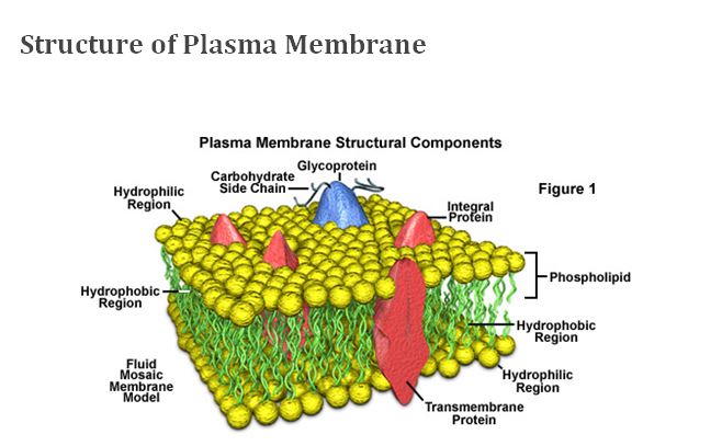
According to the accepted current theory, known as the fluid mosaic model, the plasma membrane is composed of a double layer (bilayer) of lipids, oily substances found in all cells (see Figure 1). Most of the lipids in the bilayer can be more precisely described as phospholipids, that is, lipids that feature a phosphate group at one end of each molecule. Phospholipids are characteristically hydrophilic (“water-loving”) at their phosphate ends and hydrophobic (“water-fearing”) along their lipid tail regions. In each layer of a plasma membrane, the hydrophobic lipid tails are oriented inwards and the hydrophilic phosphate groups are aligned so they face outwards, either toward the aqueous cytosol of the cell or the outside environment. Phospholipids tend to spontaneously aggregate by this mechanism whenever they are exposed to water.
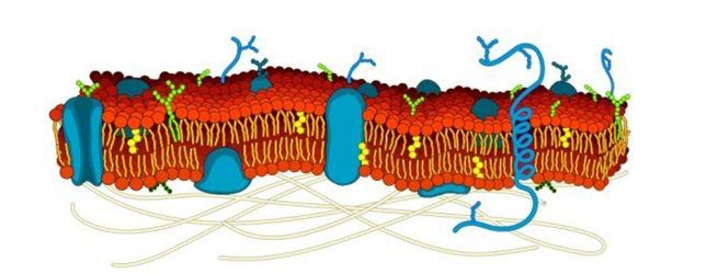
Within the phospholipid bilayer of the plasma membrane, many diverse proteins are embedded, while other proteins simply adhere to the surfaces of the bilayer. Some of these proteins, primarily those that are at least partially exposed on the external side of the membrane, have carbohydrates attached to their outer surfaces and are, therefore, referred to as glycoproteins. The positioning of proteins along the plasma membrane is related in part to the organization of the filaments that comprise the cytoskeleton, which help anchor them in place. The arrangement of proteins also involves the hydrophobic and hydrophilic regions found on the surfaces of the proteins: hydrophobic regions associate with the hydrophobic interior of the plasma membrane and hydrophilic regions extend past the surface of the membrane into either the inside of the cell or the outer environment.
Plasma membrane proteins function in several different ways. Many of the proteins play a role in the selective transport of certain substances across the phospholipid bilayer, either acting as channels or active transport molecules. Others function as receptors, which bind information-providing molecules, such as hormones, and transmit corresponding signals based on the obtained information to the interior of the cell. Membrane proteins may also exhibit enzymatic activity, catalyzing various reactions related to the plasma membrane.
Since the 1970s, the plasma membrane has been frequently described as a fluid mosaic, which is reflective of the discovery that oftentimes the lipid molecules in the bilayer can move about in the plane of the membrane. However, depending upon a number of factors, including the exact composition of the bilayer and temperature, plasma membranes can undergo phase transitions which render their molecules less dynamic and produce a more gel-like or nearly solid state. Cells are able to regulate the fluidity of their plasma membranes to meet their particular needs by synthesizing more of certain types of molecules, such as those with specific kinds of bonds that keep them fluid at lower temperatures. The presence of cholesterol and glycolipids, which are found in most cell membranes, can also affect molecular dynamics and inhibit phase transitions.
In prokaryotes and plants, the plasma membrane is an inner layer of protection since a rigid cell wall forms the outside boundary for their cells. The cell wall has pores that allow materials to enter and leave the cell, but they are not very selective about what passes through. The plasma membrane, which lines the cell wall, provides the final filter between the cell interior and the environment.
Eukaryotic animal cells are generally thought to have descended from prokaryotes that lost their cell walls. With only the flexible plasma membrane left to enclose them, these primordial creatures would have been able to expand in size and complexity. Eukaryotic cells are generally ten times larger than prokaryotic cells and have membranes enclosing interior components, the organelles. Like the exterior plasma membrane, these membranes also regulate the flow of materials, allowing the cell to segregate its chemical functions into discrete internal compartments.
Like all other cellular membranes, the plasma membrane consists of both lipids and proteins. The fundamental structure of the membrane is the phospholipid bilayer, which forms a stable barrier between two aqueous compartments. In the case of the plasma membrane, these compartments are the inside and the outside of the cell. Proteins embedded within the phospholipid bilayer carry out the specific functions of the plasma membrane, including selective transport of molecules and cell-cell recognition.
Functions of Plasma Membrane
-
- The plasma membrane functions as a physical barrier between the external environment and the inner cell organelles.
-
- The plasma membrane is a selectively permeable membrane, which permits the movement of only certain molecules both in and out of the cell.
-
- The plasma membranes play an important role in both the endocytosis and exocytosis processes.
-
- The plasma membrane also functions by facilitating communication and signalling between the cells.
-
- The plasma membrane plays a vital role in anchoring the cytoskeleton to provide shape to the cell and also maintain the cell potential.
Facts about Plasma Membrane
Both cell membrane and plasma membrane are often confused because of the similarity in words. But these two are the protective organelles of the cell and are very much different in their structure, composition and functions. The cell membrane is a type of plasma membrane and is not always the outermost layer of the cell.
For more information refer: Different Models of Plasma Membrane
This article concludes the introduction to plasma membranes, their facts and their importance. To know more about plasma membranes, their structure, functions,
The components of the plasma membrane
| Component | Location |
| Phospholipids | Main fabric of the membrane |
| Cholesterol | Tucked between the hydrophobic tails of the membrane phospholipids |
| Integral proteins | Embedded in the phospholipid bilayer; may or may not extend through both layers |
| Peripheral proteins | On the inner or outer surface of the phospholipid bilayer, but not embedded in its hydrophobic core |
| Carbohydrates | Attached to proteins or lipids on the extracellular side of the membrane (forming glycoproteins and glycolipids) |
Different models of Plasma membrane
In eukaryotic cells, the plasma membrane surrounds a cytoplasm filled with ribosomes and organelles. Organelles are structures that are themselves encased in membranes. Some organelles (nuclei, mitochondria, chloroplasts) are even surrounded by double membranes. All cellular membranes are composed of two layers of phospholipids embedded with proteins. All are selectively permeable (semi-permeable), allowing only certain substances to cross the membrane. The unique functions of cellular membranes are due to their different phospholipid and protein compositions. Decades of research have revealed these functions.
Various models of Plasma membrane:
-
- The four historical models of Plasma Membrane are:
-
- 1. Lipid and Lipid Bilayer Model (1925-26)
-
- 2. Dannelli Model. (1935)
-
- 3. Unit Membrane Model (Protein-Lipid Bilayer-Protein) (1953)
-
- 4. Fluid Mosaic Model (1972)
Lipid and Lipid Bilayer Model-
Lipid Bilayer Definition
A lipid bilayer is a biological membrane consisting of two layers of lipid molecules. Each lipid molecule, or phospholipid, contains a hydrophilic head and a hydrophobic tail. The tail regions, being repelled by water and slightly attracted to each other, congregate together. This exposes the head regions to the outside, creating a barrier between two bodies of water. A lipid bilayer is the foundational part of all cellular membranes, typically completed with species-specific integral proteins and other functional aspects.
A lipid bilayer functions through the actions of polarity. The inside of the lipid bilayer is non-polar, while the heads are polar molecules and create hydrogen bonds with other polar molecules. This also means that polar molecules like water and ions cannot as easily cross through the nonpolar tail region of the lipid bilayer. The cellular membranes of most organisms are created with lipid bilayer, as well as the nuclear membrane and various organelle membranes. The various functions of these membrane are then specified with a variety of proteins which allow or disallow certain substances to cross the membrane. In doing so, cells and individual organelles can create an ideal environment for biochemical reactions to occur, allowing them to stay in homeostasis.
Structure of the Lipid Bilayer
A lipid bilayer consists of two sheets of amphiphilic phospholipids, as seen in the image below. Amphiphilic describes a molecule which is part hydrophobic, part hydrophilic. There is often phosphorus atoms in the heads of the molecules, giving the heads polarity. The tails of the molecules are nonpolar and hydrophobic. In the image below, the polar parts of the molecules are marked in red.
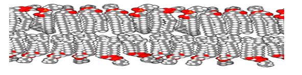
As seen in the animation, the molecules are not stuck rigidly in place. In a single sheet, the molecules are actively moving around and past each other. In fact, a better analogy is that of people crammed in an elevator. They mostly stay put, but can slide past one another if someone needs to get off the elevator and is standing in the back. Put two of these layers together, and you have a lipid bilayer.
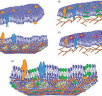
In living systems a lipid bilayer is never by itself. It is associated with a number of surface and integral proteins, as well as extracellular and intracellular elements that have specific functions in the cell. An encompassing model of the entire cellular membrane is the fluid mosaic model, which assumes that proteins within the lipid bilayer act as icebergs within the sea, drifting around but not bound to anything. The specific properties of the protein and of the lipid bilayer keep them bound within the layers, but not stationary. This can be seen in the image below.
Function of the Lipid Bilayer
A lipid bilayer serves many functions within unicellular organism and multicellular organisms alike. Regardless if a cell is living freely in pond water or confined in your body serving a function, it needs to maintain different conditions for the various reactions it needs to conduct to survive. In all applications, the lipid bilayer acts as the filter between the inside and outside. However, depending on the conditions the exact functions of the lipid bilayer can change.
Imagine two cells, one in the ocean and one in a pond. Pond water is fresh, whereas the ocean water contains many dissolved salts. In the pond, water will want to move into the more hypertonic, or saltier, cell. In the ocean, the salts in the water will draw water out of the cell. These two differing situations show how important the proteins in a lipid bilayer are. While each bilayer stops the ions and slows the movement of water, it can only hold back a certain pressure. Water will continually leach into or out of the cell. Different types of organisms have different strategies for dealing with water loss, most depending on proteins within the lipid bilayer or extracellular support structures (cell walls) to help mitigate water and ions appropriately.
Of these membrane proteins ion pumps, ion channels, and aquaporins. Ion pumps rely on cellular energy sources (e.g. ATP) to actively move unwanted ions across a lipid bilayer. Ion channels, on the other hand, respond to a signal (electrical or chemical) and open accordingly. Aquaporins are a type of ion channel allowing larger quantities of water to pass through the membrane at the appropriate time.
The lipid bilayer and its associated proteins provide another function for cells, in the way of cellular signaling. They can be involved in a number of ways. In signal transduction, a signal is passed through the lipid bilayer using a series of integral and surface proteins, creating a reaction internally. Lipid bilayers are also directly involved in the transmission of nerve impulses. When a nerve impulse reaches the end of a nerve, called the synapse, it sends a signal for special vesicles to fuse with the lipid bilayer of the cell membrane. The vesicles, filled with neurotransmitter molecules, release their contents upon fusing. This sends the neurotransmitter across the synaptic cleft, where the next nerve cell can receive it. On this nerve cell, the binding of the neurotransmitter to special proteins causes the formation of an electrical action potential, which moves as an electrical wave down the lipid bilayer.
A further function of the lipid bilayer is of cellular rigidity and support. The makeup of the lipid bilayer is such that at different temperatures and compositions, it acts different. According to the species and environment it lives in (hot, cold, etc.), the lipid bilayer will be made from different kinds and types of lipids. For example, humans produce a lipid called cholesterol, which influences the stiffness of the cellular membrane. With more cholesterol between the other lipid molecules in the bilayer, the entire structure becomes more rigid. This becomes a problem when there is too much cholesterol, as cells can no longer bend and flex as they are meant to. In humans and other animals this lead to tears in the walls of arteries, which are under immense pressure from the heart. If these arteries tear, you can bleed out internally.
Finally, in a number of species the lipid bilayer is involved in the processes of endocytosis and exocytosis. Taking in food and excreting substances, respectively, are the simple definitions of these terms. During these events, the lipid bilayer is folded (or unfolded) to take in (or excrete) substances. While there are several types of endocytosis, phagocytosis is the act of enveloping a prey or food item by folding the lipid bilayer around it and forming an internal vesicle in which the item can be digested. This method is practiced by a number of unicellular species in feeding.
My Other Post-