Chromosome
Contents for Tables:-
| S.R.No. | Contents | Page No. |
| 1 | Acknowledgement | 3-3 |
| 2 | Declaration | 4-4 |
| 3 | Introduction | 5-5 |
| 4 | Chromosome in Prokaryotic Cell | 6-7 |
| 5 | Chromosome in eukaryotic Cell | 7-9 |
| 6 | Chromosome and Plasmids | 9-11 |
| 7 | Giant Chromosome | 11-13 |
| 8 | Chromosome Meaning and Discovery | 13-14 |
| 9 | Chromosome Structure | 15-19 |
| 10 | Type of Chromosome | 20-37 |
| 11 | Function of Chromosome | 37-39 |
Introduction
A chromosome is a package of DNA with part or all of the genetic material of an organism. In most chromosomes, the very long thin DNA fibers are coated with nucleosome forming packaging proteins; in eukaryotic cells the most important of these proteins are the histones. These proteins, aided by chaperone proteins, bind to and condense the DNA molecule to maintain its integrity. These chromosomes display a complex three-dimensional structure, which plays a significant role in transcriptional regulation.
Chromosomes are normally visible under a light microscope only during the metaphase of cell division (where all chromosomes are aligned in the center of the cell in their condensed form). Before this happens, each chromosome is duplicated (S phase), and both copies are joined by a centromere, resulting either in an X-shaped structure (pictured above), if the centromere is located equatorially, or a two-arm structure, if the centromere is located distally. The joined copies are now called sister chromatids. During metaphase the X-shaped structure is called a metaphase chromosome, which is highly condensed and thus easiest to distinguish and study. In animal cells, chromosomes reach their highest compaction level in anaphase during chromosome segregation.
Chromosomal recombination during meiosis and subsequent sexual reproduction play a significant role in genetic diversity. If these structures are manipulated incorrectly, through processes known as chromosomal instability and translocation, the cell may undergo mitotic catastrophe. Usually, this will make the cell initiate apoptosis leading to its own death, but sometimes mutations in the cell hamper this process and thus cause progression of cancer.
Some use the term chromosome in a wider sense, to refer to the individualized portions of chromatin in cells, either visible or not under light microscopy. Others use the concept in a narrower sense, to refer to the individualized portions of chromatin during cell division, visible under light microscopy due to high condensation.
Chromosomes
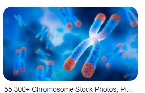
Chromosomes are present in the nucleus and they carry genetic information. They play an important role in cell division, heredity, variation, growth and repair, etc. The term chromosome was coined by W. Waldeyer in 1883 to describe darkly stained material in the nucleus.
Chromosomes in prokaryotic cells
Prokaryotic cells mostly have a single circular chromosome. Some bacteria have linear chromosomes also. They are present in the cytoplasm as the nucleus is absent. DNA is not scattered all around in the cell but is present in the nucleoid region. The DNA makes large loops and is held by proteins. The organisation of chromosomes in prokaryotes is less complex compared to eukaryotes.
Bacteria often get a bad rap: they’re described as unsafe “bugs” that cause disease. Although some types of bacteria do cause disease (as you know if you’ve ever been prescribed antibiotics), many other are harmless, or even beneficial.
Bacteria are classified as prokaryotes, along with another group of single-celled organisms, the archaea. Prokaryotes are tiny, but in a very real sense, they dominate the Earth. They live nearly everywhere – on every surface, on land and in water, and even inside of our bodies.
To emphasize that last point: you probably have about the same number of prokaryotic cells in your body as human cells! That may sound gross, but many of our prokaryotic “sidekicks” play important roles in keeping us healthy.
In this article, we’ll look at what prokaryotes are and what exactly makes them different from eukaryotes (such as you, a houseplant, or a fungus). Then, we’ll take a closer look at the structures these efficient, omnipresent little organisms use to survive.
Chromosomes in eukaryotic cells
Eukaryotic chromosomes contain a linear DNA double helix associated with proteins. Chromosomes appear as long thin threads at the interphase called chromatin. DNA is wrapped around the histone octamer to form a nucleosome, which is the repeating unit of chromatin thread. Chromatin fibres coil and condense further to form chromosomes at the metaphase. The structure of chromosomes is mostly studied at mitotic metaphase.
Chromosome number, size, shape, etc. varies in different organisms. Each diploid cell contains two sets of homologous chromosomes.
Eukaryotic cells are defined as cells containing organized nucleus and organelles which are enveloped by membrane-bound organelles. Examples of eukaryotic cells are plants, animals, protists, fungi. Their genetic material is organized in chromosomes. Golgi apparatus, Mitochondria, Ribosomes, Nucleus are parts of Eukaryotic Cells. Let’s learn about the parts of eukaryotic cells in detail.
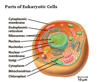
Cytoplasmic Membrane
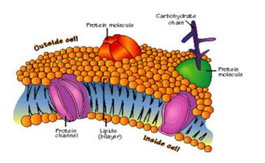
Description: It is also called plasma membrane or cell membrane. The plasma membrane is a semi-permeable membrane that separates the inside of a cell from the outside.
Structure and Composition: In eukaryotic cells, the plasma membrane consists of proteins, carbohydrates and two layers of phospholipids (i.e. lipid with a phosphate group). These phospholipids are arranged as follows:
-
- The polar, hydrophilic (water-loving) heads face the outside and inside of the cell. These heads interact with the aqueous environment outside and within a cell.
-
- The non-polar, hydrophobic (water-repelling) tails are sandwiched between the heads and are protected from the aqueous environments.
Scientists Singer and Nicolson described the structure of the phospholipid bilayer as the ‘Fluid Mosaic Model’. The reason is that the bi-layer looks like a mosaic and has a semi-fluid nature that allows lateral movement of proteins within the bilayer.
Chromosome and plasmids
Most prokaryotes have a single circular chromosome, and thus a single copy of their genetic material. Eukaryotes like humans, in contrast, tend to have multiple rod-shaped chromosomes and two copies of their genetic material (on homologous chromosomes).
Also, prokaryotic genomes are generally much smaller than eukaryotic genomes. For instance, the E. coli genome is less than half the size of the genome of yeast (a simple, single-celled eukaryote), and almost times smaller than the human genome.
By definition, prokaryotes lack a membrane-bound nucleus to hold their chromosomes. Instead, the chromosome of a prokaryote is found in a part of the cytoplasm called a nucleoid.
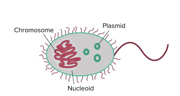
Prokaryotes generally have a single circular chromosome that occupies a region of the cytoplasm called a nucleoid. They also may contain small rings of double-stranded extra-chromosomal DNA called plasmids.
In addition to the chromosome, many prokaryotes have plasmids, which are small rings of double-stranded extra-chromosomal (“outside the chromosome”) DNA. Plasmids carry a small number of non-essential genes and are copied independently of the chromosome inside the cell. They can be transferred to other prokaryotes in a population, sometimes spreading genes that are beneficial to survival.
For instance, some plasmids carry genes that make bacteria resistant to antibiotics. (These genes are called R genes.) When the plasmids carrying R genes are exchanged in a population, they can quickly make the population resistant to antibiotic drugs. While beneficial to the bacteria, this process can make it difficult for doctors to treat harmful bacterial infections.
Giant Chromosomes
Polytene chromosome
-
- Balbiani first discovered a structure in the nuclei of secretory glands of midges
- Painter, Heitz and Bauer, rediscovered them in the salivary gland of Drosophila and recognised them as a chromosome
- Also known as Salivary gland chromosome
- These are called polytene by Kollar due to the presence of many chromonemata in them
- These are present in some cells of the larvae of Dipteran insects
- These are very large due to the presence of high DNA content
- The polytene chromosome of Drosophila’s salivary gland has 1000 DNA molecules Chironomus has 1600 DNA molecules in its each polytene chromosome
- There is a series of alternating dark and clear bands called interband
- Chromosome puffs or Balbiani rings are present, which are the swelling of bands due to DNA unfolding into open loops. These are the region of the intense transcription or mRNA formation
-
- Lampbrush chromosome
- Balbiani first discovered a structure in the nuclei of secretory glands of midges
-
- First discovered in the oocytes of salamander
-
- The name is given due to its resemblance with a brush that is used for cleaning lamp, glass chimneys, etc.
-
- They occur at the diplotene stage of oocytes in vertebrates and invertebrates
-
- Lampbrush chromosomes are also found in the spermatocytes of many animals and also found in the giant nucleus of an algae Acetabularia
-
- They are present as a bivalent with 4 chromatids
-
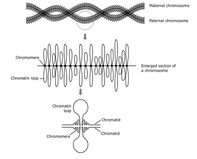
- Chromosomal axis is formed from highly condensed chromatin and lateral loops extend from the row of chromomeres
-
- Lateral loops of DNA are always symmetrical and formed due to intense RNA synthesis
-
- In the oocytes of salamander, there are 10,000 loops present per haploid set of chromosomes
-
- The centromere doesn’t bear any loops
Chromosome Meaning and Discovery
Chromosome means ‘coloured body’, that refers to its staining ability by certain dyes.
Karl Nägeli in 1842, first observed the rod-like structure present in the nucleus of the plant cell.
W. Waldeyer in 1888 coined the term ‘chromosome’.
Walter Sutton and Theodor Boveri in 1902 suggested that chromosomes are the physical carrier of genes in the eukaryotic cells.
The number of chromosomes in any species is constant for all the cells. The number of chromosomes in gametes (e.g. sperms, egg) is half of the somatic cell and known as a haploid set of chromosomes, which is the result of meiosis during sexual reproduction. Chromosome number is preserved in the mitotic division of somatic cells, which is required for an organism to grow, repair and regenerate.
Chromosome number varies in different species. A nematode species contains only 2 chromosomes in a cell, whereas a protozoan species contains as much as 1600 chromosomes in the cell. Most of the plant and animal species contain 8 to 50 number of chromosomes in its somatic cell. The number of chromosomes does not reflect the complexity of a species. A human cell contains total 23 pair of chromosomes (2n, total 23×2=46), of which 22 are autosomes and 1 sex chromosome.
Karyotyping is a technique to study the structure of chromosomes present in a species. Chromosomes are isolated, stained and photographed. This technique is useful in finding out any chromosomal abnormalities.
Chromosome Structure
The chemical composition of a chromosome is histone proteins and DNA. Each cell has a pair of each kind of chromosome known as a homologous chromosome. Chromosomes are made up of chromatin, which contains a single molecule of DNA and associated proteins. Each chromosome contains hundreds and thousands of genes that can precisely code for several proteins in the cell. Structure of a chromosome can be best seen during cell division.
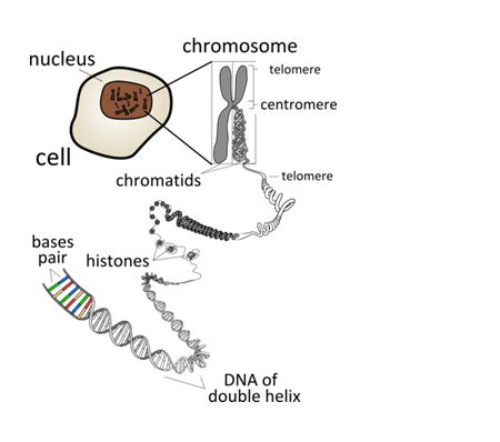
Main parts of chromosomes are:
-
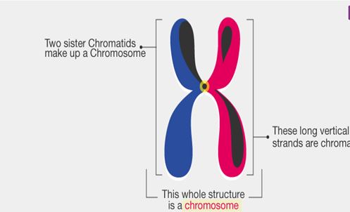
- Chromatid: Each chromosome has two symmetrical structures called chromatids or sister chromatids which is visible in mitotic metaphase.
- Each chromatid contains a single DNA molecule
-
- At the anaphase of mitotic cell division, sister chromatids separate and migrate to opposite poles
- Chromatid: Each chromosome has two symmetrical structures called chromatids or sister chromatids which is visible in mitotic metaphase.
-
- Centromere and kinetochore: Sister chromatids are joined by the centromere.
- Spindle fibres during cell division are attached at the centromere
- The number and position of the centromere differs in different chromosomes
- The centromere is called primary constriction
- Centromere divides the chromosome into two parts, the shorter arm is known as ‘p’ arm and the longer arm is known as ‘q’ arm.
- The centromere contains a disc-shaped kinetochore, which has specific DNA sequence with special proteins bound to them
-
- The kinetochore provides the centre for polymerisation of tubulin proteins and assembly of microtubules
- Centromere and kinetochore: Sister chromatids are joined by the centromere.
-
- Secondary constriction and nucleolar organisers: Other than centromere, chromosomes possess secondary constrictions.
- Secondary constrictions can be identified from centromere at anaphase because there is bending only at the centromere (primary constriction)
-
- Secondary constrictions, which contain genes to form nucleoli are known as the nucleolar organiser
- Secondary constriction and nucleolar organisers: Other than centromere, chromosomes possess secondary constrictions.
-
- Telomere: Terminal part of a chromosome is known as a telomere.
-
- Telomeres are polar, which prevents the fusion of chromosomal segments
-
- Telomere: Terminal part of a chromosome is known as a telomere.
-
- Satellite: It is an elongated segment that is sometimes present on a chromosome at the secondary constriction.
-
- The chromosomes with satellite are known as sat-chromosome
-
- Satellite: It is an elongated segment that is sometimes present on a chromosome at the secondary constriction.
-
- Chromatin: Chromosome is made up of chromatin. Chromatin is made up of DNA, RNA and proteins. At interphase, chromosomes are visible as thin chromatin fibres present in the nucleoplasm. During cell division, the chromatin fibres condense and chromosomes are visible with distinct features.
- The darkly stained, condensed region of chromatin is known as heterochromatin. It contains tightly packed DNA, which is genetically inactive
- The light stained, diffused region of chromatin is known as euchromatin. It contains genetically active and loosely packed DNA
- At prophase, the chromosomal material is visible as thin filaments known as chromonemata
-
- At interphase, bead-like structures are visible, which are an accumulation of chromatin material called chromomere. Chromatin with chromomere looks like a necklace with beads
- Chromatin: Chromosome is made up of chromatin. Chromatin is made up of DNA, RNA and proteins. At interphase, chromosomes are visible as thin chromatin fibres present in the nucleoplasm. During cell division, the chromatin fibres condense and chromosomes are visible with distinct features.
Labelled diagram of chromosome is given below.
My Other Post-

Если ваш старый пульт сломался или вы просто хотите обновить его до более современного и функционального варианта, то самое время универсальный пульт управления купить, который идеально подойдет для ваших потребностей и обеспечит комфортное управление вашим телевизором.
Приобретая телевизор, важно подумать о покупке пульта для телевизора. Это связано с тем, что пульт для телевизора является неотъемлемой частью телевизионной системы . С помощью пульта можно выполнять различные функции, такие как переключение каналов и регулировка громкости .
При приобретении пульта для телевизора следует рассмотреть несколько ключевых аспектов. Одним из них является совместимость пульта с телевизором . Другим важным фактором является дизайн и эргономика пульта .
## Раздел 2: Типы пультов для телевизоров
В продаже имеются разные виды пультов для телевизоров, имеющие различные особенности. Один из наиболее популярных типов пультов – это пульт с инфракрасной связью. Этот тип пульта работает за счет передачи инфракрасного сигнала на телевизор.
Другим типом пульта является пульт с радиочастотным сигналом . Этот тип пульта работает за счет передачи радиосигнала на телевизор. Также имеются пульты с блютуз-соединением, которые ermogняют управление телевизором через мобильное устройство .
## Раздел 3: Выбор пульта для телевизора
При выборе пульта для телевизора необходимо учитывать несколько факторов . Одним из них является совместимость пульта с телевизором . Другим аспектом является дизайн и эргономичность пульта.
Помимо этого, важно рассмотреть функциональные возможности пульта. Некоторые пульты обладают дополнительными возможностями, такими как управление другими гаджетами . При приобретении пульта следует изучить отзывы других владельцев.
## Раздел 4: Заключение
В итоге, приобретение пульта для телевизора является ключевым шагом в создании комфортной телевизионной системы . При выборе пульта необходимо учитывать несколько факторов, включая совместимость, дизайн и функциональность . С помощью правильного пульта можно повысить уровень комфорта при просмотре телевизора .
Если ваш старый пульт сломался или вы просто хотите обновить его до более современного и функционального варианта, то самое время купить пульт интернет магазин, который идеально подойдет для ваших потребностей и обеспечит комфортное управление вашим телевизором.
Приобретая телевизор, важно подумать о покупке пульта для телевизора. Это связано с тем, что пульт для телевизора играет ключевую роль в управлении телевизором. Благодаря пульту можно легко переключать каналы, регулировать громкость и настраивать другие параметры телевизора .
При приобретении пульта для телевизора следует рассмотреть несколько ключевых аспектов. Одним из них является совместимость пульта с телевизором . Другим аспектом является дизайн и эргономичность пульта.
## Раздел 2: Типы пультов для телевизоров
Существует несколько типов пультов для телевизоров, каждый из которых имеет свои уникальные характеристики . Одним из наиболее распространенных типов является пульт с инфракрасным сигналом . Этот тип пульта работает за счет передачи инфракрасного сигнала на телевизор.
Другой тип пульта – это пульт с радиочастотной связью. Этот тип пульта передает сигнал на телевизор с помощью радиоволн . Кроме того, существуют пульты с блютуз-соединением, которые позволяют управлять телевизором с помощью смартфона .
## Раздел 3: Выбор пульта для телевизора
При выборе пульта для телевизора необходимо учитывать несколько факторов . Одним из них является совместимость пульта с телевизором . Другим моментом, на который стоит обратить внимание, является дизайн и удобство использования пульта .
Кроме того, важно учитывать функциональность пульта . Некоторые пульты имеют расширенные функции, такие как управление другими приборами. При выборе пульта необходимо прочитать отзывы других пользователей .
## Раздел 4: Заключение
В заключении, покупка пульта для телевизора является важным этапом в настройке домашнего кинотеатра . При выборе пульта необходимо учитывать несколько факторов, включая совместимость, дизайн и функциональность . С помощью правильного пульта можно повысить уровень комфорта при просмотре телевизора .
На современном рынке существует множество поставщиков крепежа, предлагающих широкий ассортимент изделий.
Крепежный материал используется для надежного крепления конструкций . Это обусловлено его способностью надежно соединять различные элементы, обеспечивая прочность и долговечность конечного продукта Крепление деталей с помощью крепежа гарантирует надежность конструкции . Использование качественного крепежа имеет решающее значение для предотвращения поломок и обеспечения безопасности эксплуатации Использование качественного крепежа предотвращает поломки .
Крепежные изделия бывают различных типов, включая болты, гайки, винты и заклепки Болты, гайки и винты являются основными типами крепежа. Каждый тип имеет свои особенности и области применения Винты и заклепки применяются для крепления деталей в различных конструкциях. Правильный выбор крепежа зависит от конкретных требований проекта и свойств материалов Тип крепежа выбирается в зависимости от требований проекта.
Болты и гайки являются одними из наиболее распространенных типов крепежа Болты и гайки используются для создания прочных соединений . Они широко используются в строительстве, машиностроении и других отраслях Болты и гайки применяются в различных отраслях промышленности . Болты и гайки бывают различных размеров и типов Болты и гайки имеют разные характеристики и свойства.
Винты и заклепки также широко используются для крепления деталей Винты и заклепки используются для крепления деталей в различных конструкциях . Винты могут быть саморезирующими или обычными Винты могут быть сделаны из различных материалов . Заклепки используются для соединения деталей в различных отраслях Заклепки применяются в строительстве и производстве мебели .
Крепеж используется в различных отраслях промышленности, включая строительство, машиностроение и производство мебели Крепеж используется для соединения деталей в различных конструкциях. В строительстве крепеж используется для соединения деталей зданий и сооружений Крепеж используется для соединения деталей зданий . В машиностроении крепеж используется для создания прочных соединений в механизмах и агрегатах Крепеж используется для создания прочных соединений в машинах .
Крепеж также используется в производстве мебели для соединения деталей и обеспечения прочности конструкции Крепеж используется для соединения деталей мебели . Правильный выбор крепежа зависит от типа материала и требований проекта Тип крепежа выбирается в зависимости от требований проекта. Использование качественного крепежа имеет решающее значение для предотвращения поломок и обеспечения безопасности эксплуатации Правильный выбор крепежа обеспечивает надежную работу оборудования.
Будущее крепежа связано с развитием новых материалов и технологий Будущее крепежа зависит от технологических достижений . Использование композитных материалов и нанотехнологий может значительно улучшить свойства крепежа Использование композитных материалов улучшит свойства крепежа . Кроме того, развитие робототехники и автоматизации может повысить эффективность и точность сборки и крепления Робототехника и автоматизация меняют подход к крепежу.
Развитие 3D-печати и аддитивных технологий также может оказать существенное влияние на производство и применение крепежа 3D-печать и аддитивные технологии меняют подход к проектированию и производству крепежа. В будущем можно ожидать появления новых типов крепежа, которые будут обладать улучшенными свойствами и характеристиками В будущем появятся новые типы крепежа . Это, в свою очередь, будет способствовать развитию новых технологий и отраслей Новые технологии и отрасли будут тесно связаны с будущим крепежа.
На современном рынке существует множество поставщиков крепежа, предлагающих широкий ассортимент изделий.
Крепежные изделия являются основой для создания прочных соединений. Это обусловлено его способностью надежно соединять различные элементы, обеспечивая прочность и долговечность конечного продукта Крепление деталей с помощью крепежа гарантирует надежность конструкции . Использование качественного крепежа имеет решающее значение для предотвращения поломок и обеспечения безопасности эксплуатации Качественный крепеж гарантирует безопасность эксплуатации .
Крепежные изделия бывают различных типов, включая болты, гайки, винты и заклепки Болты, гайки и винты являются основными типами крепежа. Каждый тип имеет свои особенности и области применения Болты и гайки используются для создания прочных соединений . Правильный выбор крепежа зависит от конкретных требований проекта и свойств материалов Выбор крепежа осуществляется с учетом нагрузки и типа соединения .
Болты и гайки являются одними из наиболее распространенных типов крепежа Болты и гайки представляют собой надежный крепеж. Они широко используются в строительстве, машиностроении и других отраслях Болты и гайки обеспечивают надежное крепление конструкций. Болты и гайки бывают различных размеров и типов Болты и гайки имеют разные характеристики и свойства.
Винты и заклепки также широко используются для крепления деталей Винты и заклепки используются для крепления деталей в различных конструкциях . Винты могут быть саморезирующими или обычными Винты могут быть сделаны из различных материалов . Заклепки используются для соединения деталей в различных отраслях Заклепки применяются в строительстве и производстве мебели .
Крепеж используется в различных отраслях промышленности, включая строительство, машиностроение и производство мебели Крепеж используется для соединения деталей в различных конструкциях. В строительстве крепеж используется для соединения деталей зданий и сооружений Крепеж обеспечивает надежное крепление кровли и фасадов. В машиностроении крепеж используется для создания прочных соединений в механизмах и агрегатах Крепеж обеспечивает надежную работу оборудования.
Крепеж также используется в производстве мебели для соединения деталей и обеспечения прочности конструкции Крепеж используется для соединения деталей мебели . Правильный выбор крепежа зависит от типа материала и требований проекта Правильный выбор крепежа зависит от типа материала . Использование качественного крепежа имеет решающее значение для предотвращения поломок и обеспечения безопасности эксплуатации Использование качественного крепежа предотвращает поломки .
Будущее крепежа связано с развитием новых материалов и технологий Будущее крепежа зависит от технологических достижений . Использование композитных материалов и нанотехнологий может значительно улучшить свойства крепежа Нанотехнологии могут повысить прочность и долговечность крепежа . Кроме того, развитие робототехники и автоматизации может повысить эффективность и точность сборки и крепления Развитие робототехники улучшит эффективность сборки .
Развитие 3D-печати и аддитивных технологий также может оказать существенное влияние на производство и применение крепежа 3D-печать изменит подход к производству крепежа . В будущем можно ожидать появления новых типов крепежа, которые будут обладать улучшенными свойствами и характеристиками В будущем появятся новые типы крепежа . Это, в свою очередь, будет способствовать развитию новых технологий и отраслей Новые технологии и отрасли будут тесно связаны с будущим крепежа.
Для тех, кто ищет выгодные предложения на ткани, купить ткани оптом в москве дешево предлагает широкий выбор тканей по разумным ценам.
как найти оптовых поставщиков тканей является первым шагом к успеху. Это связано с тем, как оптовые торговцы работают напрямую с производителями. При этом как качество тканей напрямую влияет на конечный продукт .
где оптовые покупки являются выгодными для бизнеса. Кроме того, как можно выбрать ткани разной текстуры и цвета . Это особенно важно где оптовые поставщики тканей могут предложить высококачественные материалы.
Одним из лучших мест для поиска оптовых поставщиков тканей являются онлайн-площадки . Кроме того, посещение отраслевых выставок и ярмарок может стать отличной возможностью . При этом как можно сэкономить на доставке .
Использование поисковых систем для поиска оптовых поставщиков тканей также является эффективным методом . Кроме того, как узнать о надежных поставщиках . Это особенно важно где оптовые поставщики тканей могут предложить flexible условия работы.
как качество предлагаемых тканей . Кроме того, где найти информацию о компании. При этом где найти компанию с flexible условиями.
как можно гарантировать соответствие ткани необходимым стандартам . Кроме того, где найти компанию с профессиональной поддержкой. Это особенно важно для бизнеса, который планирует долгосрочное сотрудничество .
В заключении, поиск оптового поставщика тканей требует тщательного подхода . Кроме того, где найти надежного партнера является ключом к успеху. При этом постоянный поиск новых поставщиков и мониторинг рынка также необходимы .
Будущее оптовой торговли тьмается перспективным, и где найти инновационных поставщиков. Кроме того, экологичность и устойчивость также станут ключевыми факторами в выборе оптового поставщика тканей . Это особенно важно где оптовые поставщики тканей могут предложить высококачественные материалы и flexible условия.
Чтобы освободить территорию после ремонта, вам поможет заказ вывоза строительного мусора.
важно грамотно утилизировать остатки
Чтобы освободить территорию после ремонта, вам поможет вывоз мусора строительные отходы.
строительстве
Для покупки пластиковые флаконы купить можно обратиться напрямую к производителю или крупному поставщику, что позволит экономить на покупке и обеспечить качественный продукт для различных применений.
Флакон опт — это оптимальный выбор для покупателей, которые ищут ценный и удобный продукт. Кроме того, покупка флакона в опте является выгодным решением для всех. Также Флакон опт имеет множество преимуществ, включая экономию средств и получение лучшего качества. Кроме того, покупка флакона оптом позволяет приобрести необходимый продукт в большом количестве. Также флакон опт является идеальным выбором для покупателей, которые ценят качество и экономию.
Флакон опт предлагает покупателям множество преимуществ, включая экономию средств и получение лучшего качества. Кроме того, покупка флакона оптом позволяет приобрести необходимый продукт в большом количестве. Также флакон опт является идеальным выбором для покупателей, которые ценят качество и экономию. Кроме того, покупка флакона оптом позволяет сэкономить средства и получить лучшее качество. Также флакон опт является лучшим решением для тех, кто ищет качественный и удобный продукт.
флакон опт необходимо выбирать исходя из качества и цены. Кроме того, покупка флакона в опте требует анализа качества и цены. Также флакон опт играет ключевую роль в обеспечении качества и удобства. Кроме того, покупка флакона оптом требует учета потребностей и требований покупателя. Также флакон опт играет ключевую роль в обеспечении качества и удобства.
Флакон опт является оптимальным выбором для покупателей, которые ищут ценный и удобный продукт. Кроме того, покупка флакона в опте является выгодным решением для всех. Также флакон опт широко используется среди покупателей оптовых товаров. Кроме того, покупка флакона в опте дает возможность приобрести лучший продукт по низкой цене. Также флакон опт является лучшим решением для тех, кто ищет качественный и удобный продукт.
Если намереваетесь совершить поездку из Калининграда в Зеленоградск, настоятельно советую обратить внимание на электричку Калининград Зеленоградск — это практичный и выгодный способ добраться до побережья. Протяженность маршрута от Калининграда до Зеленоградска составляет порядка 40 км, а маршрут следует мимо чудесных пейзажей и предоставляет шанс быстро оказаться у пляжей Калининградской области. Также не пропустите замок Шаакен Калининградской области — это реальная историческая сокровище региона и замечательное место для прогулок.
Если хотите почувствовать характер местной жизни, отправляйтесь на барахолку в Калининграде, где можно выбрать уникальные антикварные вещи и сувениры. Для отдыха с детьми идеально подойдут парк миниатюр в Калининграде и зоопарк Калининград — они понравятся всей семье. Детальный путеводитель по достопримечательностям и расписания транспорта можно изучить здесь зоопарк калининграда .
Для покупки полимерные флаконы можно обратиться напрямую к производителю или крупному поставщику, что позволит экономить на покупке и обеспечить качественный продукт для различных применений.
Флакон опт является идеальным решением для тех, кто ценит качество и экономию. Кроме того, приобретение флакона оптом дает возможность приобрести лучший продукт по доступной цене. Также Флакон опт имеет множество преимуществ, включая экономию средств и получение лучшего качества. Кроме того, покупка флакона в опте дает возможность приобрести лучший продукт по низкой цене. Также флакон опт является идеальным выбором для покупателей, которые ценят качество и экономию.
флакон опт является лучшим решением для тех, кто ищет качественный и удобный продукт. Кроме того, покупка флакона в опте дает возможность приобрести лучший продукт по низкой цене. Также флакон опт дает возможность приобрести лучший продукт по доступной цене. Кроме того, покупка флакона оптом позволяет приобрести необходимый продукт в большом количестве. Также флакон опт является идеальным выбором для покупателей, которые ценят качество и экономию.
флакон опт необходимо выбирать исходя из качества и цены. Кроме того, покупка флакона оптом требует тщательного рассмотрения и сравнения вариантов. Также флакон опт необходимо выбирать исходя из опыта и отзывов других покупателей. Кроме того, покупка флакона в опте требует анализа качества и цены. Также флакон опт является важным фактором при принятии решения о покупке.
флакон опт — это лучший способ удовлетворить потребности в опте. Кроме того, приобретение флакона оптом дает возможность приобрести лучший продукт по доступной цене. Также флакон опт предлагает покупателям выгодные условия и лучшее качество. Кроме того, покупка флакона оптом позволяет сэкономить средства и получить лучшее качество. Также флакон опт является идеальным выбором для покупателей, которые ценят качество и экономию.
Creez un espace cosy dans votre region avec pergola bioclimatique autoportante, parfait a tout moment de l’annee.
Verifiez aussi le mecanisme des lames pour s’assurer qu’il fonctionne correctement.
Creez un espace cosy dans votre region avec auvent bioclimatique, parfait a tout moment de l’annee.
Elle vous protege des intemperies tout en facilitant la circulation de l’air.
Yo, been playing around on win92. It’s got some interesting stuff going on. Might be worth a look if you’re bored: win92.
Если вы ищете качественные и стильные устройства для vaping, то hqd одноразки может стать вашим лучшим выбором, предлагая широкий выбор товаров и услуг по vaping, включая одноразовые устройства, различные вкусы и аксессуары.
Они представляют собой высококачественные устройства для vaping, созданные компанией HQD . Эти устройства очень удобны в использовании Поскольку они не требуют никакого обслуживания или настройки . HQD одноразки также очень безопасны Они изготовлены из высококачественных материалов .
HQD одноразки имеют широкий выбор вкусов Каждый вкус уникален и создан для удовлетворения разных предпочтений. Эти устройства также очень компактны Их можно легко положить в карман или сумку . HQD одноразки идеально подходят для тех, кто хочет попробовать вейпинг Поскольку они очень просты в использовании .
HQD одноразки имеют много преимуществ Они не требуют никакого обслуживания или настройки . Эти устройства также очень эффективны Поскольку они имеют высококачественные батареи . HQD одноразки также очень безопасны Они соответствуют всем необходимым стандартам безопасности.
HQD одноразки идеально подходят для тех, кто хочет бросить курить Они могут помочь улучшить здоровье. Эти устройства также очень универсальны Они очень просты в использовании. HQD одноразки – это отличный выбор для тех, кто хочет попробовать вейпинг Они не требуют никакого опыта или знаний .
HQD одноразки имеют широкий выбор вкусов Вкусы варьируются от классических табачных до экзотических фруктовых . Эти устройства также очень компактны Их можно легко положить в карман или сумку . HQD одноразки идеально подходят для тех, кто хочет попробовать разные вкусы Они являются отличным введением в мир вейпинга.
HQD одноразки также имеют разные уровни никотина Каждый уровень никотина уникален и создан для удовлетворения разных предпочтений. Эти устройства также очень экономичны Они могут быть использованы в течение долгого времени. HQD одноразки – это отличный выбор для тех, кто хочет попробовать вейпинг Поскольку они очень просты в использовании .
HQD одноразки – это популярный выбор среди любителей вейпинга Они предлагают уникальный опыт вкуса и расслабления. Эти устройства очень удобны в использовании Поскольку они не требуют никакого обслуживания или настройки . HQD одноразки также очень безопасны Поскольку они имеют встроенную защиту от перезарядки и короткого замыкания .
HQD одноразки идеально подходят для тех, кто хочет попробовать вейпинг Они не требуют никакого опыта или знаний . Эти устройства также очень универсальны Они очень просты в использовании. HQD одноразки – это отличный выбор для тех, кто хочет попробовать вейпинг Они являются отличным введением в мир вейпинга.
Если вы ищете качественные и стильные устройства для vaping, то hqd купить в москве с доставкой может стать вашим лучшим выбором, предлагая широкий выбор товаров и услуг по vaping, включая одноразовые устройства, различные вкусы и аксессуары.
Они предлагают уникальный опыт вкуса и расслабления. Эти устройства очень удобны в использовании Их можно использовать сразу после покупки. HQD одноразки также очень безопасны Они соответствуют всем необходимым стандартам безопасности.
HQD одноразки имеют широкий выбор вкусов 20 различных вкусов на выбор . Эти устройства также очень компактны Их можно взять с собой куда угодно. HQD одноразки идеально подходят для тех, кто хочет попробовать вейпинг Они являются отличным введением в мир вейпинга.
HQD одноразки имеют много преимуществ Они очень доступны по цене. Эти устройства также очень эффективны Поскольку они имеют высококачественные батареи . HQD одноразки также очень безопасны Они соответствуют всем необходимым стандартам безопасности.
HQD одноразки идеально подходят для тех, кто хочет бросить курить Поскольку они могут помочь уменьшить тягу к никотину . Эти устройства также очень универсальны Они очень просты в использовании. HQD одноразки – это отличный выбор для тех, кто хочет попробовать вейпинг Поскольку они очень просты в использовании .
HQD одноразки имеют широкий выбор вкусов Более 20 различных вкусов на выбор . Эти устройства также очень компактны Их можно взять с собой куда угодно. HQD одноразки идеально подходят для тех, кто хочет попробовать разные вкусы Они являются отличным введением в мир вейпинга.
HQD одноразки также имеют разные уровни никотина От 0 до 20 мг на мл . Эти устройства также очень экономичны Они очень долговечны . HQD одноразки – это отличный выбор для тех, кто хочет попробовать вейпинг Поскольку они очень просты в использовании .
HQD одноразки – это популярный выбор среди любителей вейпинга Они представляют собой высококачественные устройства для vaping, созданные компанией HQD . Эти устройства очень удобны в использовании Поскольку они не требуют никакого обслуживания или настройки . HQD одноразки также очень безопасны Они изготовлены из высококачественных материалов .
HQD одноразки идеально подходят для тех, кто хочет попробовать вейпинг Они не требуют никакого опыта или знаний . Эти устройства также очень универсальны Они не требуют никакого специального оборудования . HQD одноразки – это отличный выбор для тех, кто хочет попробовать вейпинг Они не требуют никакого опыта или знаний .
Для крупных строительных проектов оптимальным решением будет приобретение купить щебень известняковый 40 70, что позволит сэкономить средства и обеспечить необходимое качество строительных работ.
Щебень из известняка является ключевым компонентом многих строительных смесей. Этот материал используется для создания прочных и долговечных конструкций. Известняковый щебень, купленный оптом, может существенно повлиять на сокращение затрат на стройматериалы.
Известняковый щебень в больших количествах позволяет реализовывать масштабные проекты . Известняковый щебень в оптовых партиях доступен для заказа на производственных предприятиях.
Среди преимуществ известнякового щебня стоит отметить его пожаробезопасность. Известняковый щебень характеризуется улучшенными физико-механическими свойствами . Приобретение оптом известнякового щебня дает возможность сократить расходы на логистику .
Известняковый щебень, купленный оптом, используется для дорожного строительства. Оптовая продажа известнякового щебня способствует увеличению скорости строительства .
Известняковый щебень используется в дорожном строительстве для создания прочных и долговечных покрытий . Известняковый щебень характеризуется универсальностью применения . Приобретение оптом известнякового щебня дает возможность получить щебень для реализации индивидуальных проектов .
Известняковый щебень в оптовых партиях может быть доставлен на строительную площадку . Известняковый щебень в оптовых партиях доступен для заказа через мобильные приложения.
Щебень из известняка используется в различных отраслях промышленности . Приобретение оптом известнякового щебня дает возможность сократить расходы на материалы . Известняковый щебень, купленный оптом, может быть использован для реализации инфраструктурных проектов.
Щебень известняковый, купленный оптом, может быть доставлен в любую точку страны . Приобретение оптом известнякового щебня дает возможность получить щебень для реализации индивидуальных проектов .
Для крупных строительных проектов оптимальным решением будет приобретение заказать щебень известняковый, что позволит сэкономить средства и обеспечить необходимое качество строительных работ.
Известняковый щебень широко используется в строительстве благодаря своим уникальным свойствам . Этот материал известен своей высокой прочностью и долговечностью . Применение известнякового щебня в оптовых масштабах позволяет сократить расходы на строительство .
Приобретение оптом известнякового щебня дает возможность оптимизировать строительный процесс. Оптовая продажа известнякового щебня осуществляется через официальные дилерские сети .
Основными преимуществами известнякового щебня являются его долговечность и экологичность . Известняковый щебень используется в строительстве благодаря своим техническим преимуществам. Известняковый щебень, купленный в больших количествах, может быть хранен на складе для последующего использования.
Оптовые закупки известнякового щебня осуществляются для реализации инфраструктурных проектов . Оптовая продажа известнякового щебня способствует увеличению скорости строительства .
Известняковый щебень используется в дорожном строительстве для создания прочных и долговечных покрытий . Щебень из известняка может быть использован в ландшафтном дизайне . Приобретение оптом известнякового щебня дает возможность получить щебень для реализации индивидуальных проектов .
Оптовые закупки известнякового щебня включают в себя сервисную поддержку . Известняковый щебень в оптовых партиях доступен для заказа через мобильные приложения.
Щебень из известняка используется в различных отраслях промышленности . Известняковый щебень, купленный оптом, способствует экономии средств. Известняковый щебень, купленный оптом, может быть использован для реализации инфраструктурных проектов.
Щебень известняковый, купленный оптом, может быть доставлен в любую точку страны . Известняковый щебень, купленный оптом, может быть использован для строительных и ремонтных работ.
Посетите магазин велосипедов складные сегодня и найдите велосипед своей мечты!
Магазин предлагает огромный выбор велосипедов, подходящих для разных целей и стилей?? . Это место, где покупатели могут найти не только велосипеды, но и различные аксессуары и оборудование для них. Магазин велосипедов открыт для посетителей в любое время и приглашает всех любителей велосипедов к себе .
Магазин велосипедов имеет современный дизайн и комфортную обстановку, которая позволяет клиентам чувствовать себя расслабленно . Клиенты могут попробовать велосипеды перед покупкой, чтобы убедиться, что они подходят им идеально. Магазин велосипедов имеет профессиональный инструмент для ремонта и настройки велосипедов .
Магазин велосипедов имеет разнообразный ассортимент велосипедов, включая горные, шоссейные и городские модели . Каждая модель имеет свои уникальные характеристики и особенности. Магазин велосипедов имеет прямые контракты с производителями, что позволяет предлагать конкурентные цены .
Магазин велосипедов предлагает услуги по установке и настройке аксессуаров и оборудования. Клиенты могут найти все, что им нужно для комфортного и безопасного?жения. Магазин велосипедов имеет программу лояльности для постоянных клиентов .
Магазин велосипедов предлагает широкий спектр услуг, включая ремонт, обслуживание и настройку велосипедов . Клиенты могут быть уверены в высоком качестве выполненных работ. Магазин велосипедов предлагает услуги по доставке и установке запчастей и аксессуаров.
Магазин велосипедов приглашает опытных тренеров и инструкторов для проведения занятий . Клиенты могут получить новые знания и навыки, что позволит им улучшить свое?жение. Магазин велосипедов предлагает скидки и специальные условия для членов велоклубов .
Магазин велосипедов имеет опытный персонал и высококачественное оборудование. Клиенты могут найти все, что им нужно для комфортного и безопасного?жения. Магазин велосипедов работает на рынке уже несколько лет и имеет репутацию надежного партнера .
Магазин велосипедов предлагает выгодные цены и акции, что позволяет клиентам приобретать продукцию по доступным ценам . Клиенты могут быть уверены в высоком качестве продукции и услуг. Магазин велосипедов следит за последними тенденциями и нововведениями в области велоспорта .
Посетите магазины где можно купить электровелосипед сегодня и найдите велосипед своей мечты!
Магазин велосипедов является местом, где можно найти широкий выбор велосипедов для любого возраста и уровня подготовки . Это место, где покупатели могут найти не только велосипеды, но и различные аксессуары и оборудование для них. Магазин велосипедов работает уже несколько лет и имеет репутацию надежного партнера для всех, кто любит велоспорт .
Магазин велосипедов предлагает бесплатную консультацию по выбору велосипеда для каждого клиента. Клиенты могут попробовать велосипеды перед покупкой, чтобы убедиться, что они подходят им идеально. Магазин велосипедов предлагает услуги по модернизации и кастомизации велосипедов.
Магазин велосипедов предлагает широкий выбор моделей велосипедов, начиная от детских и заканчивая профессиональными . Каждая модель имеет свои уникальные характеристики и особенности. Магазин велосипедов имеет большой sklad с запасами и может быстро удовлетворять заказы клиентов.
Магазин велосипедов предлагает услуги по установке и настройке аксессуаров и оборудования. Клиенты могут найти все, что им нужно для комфортного и безопасного?жения. Магазин велосипедов предлагает скидки и бонусы для студентов и пенсионеров.
Магазин велосипедов использует только оригинальные запчасти и высококачественное оборудование. Клиенты могут быть уверены в высоком качестве выполненных работ. Магазин велосипедов имеет большую базу данных запчастей и может быстро найти любую деталь .
Магазин велосипедов приглашает опытных тренеров и инструкторов для проведения занятий . Клиенты могут получить новые знания и навыки, что позволит им улучшить свое?жение. Магазин велосипедов участвует в организации велосоревнований и поддерживает местный велоспорт.
Магазин велосипедов имеет опытный персонал и высококачественное оборудование. Клиенты могут найти все, что им нужно для комфортного и безопасного?жения. Магазин велосипедов всегда готов помочь и предоставить консультацию по любым вопросам.
Магазин велосипедов предлагает выгодные цены и акции, что позволяет клиентам приобретать продукцию по доступным ценам . Клиенты могут быть уверены в высоком качестве продукции и услуг. Магазин велосипедов всегда готов к инновациям и улучшению своей работы.
Если вам нужно флаконы купить оптом для ваших товаров, обратите внимание на различные варианты, доступные на рынке, и выбирайте то, что лучше всего соответствует потребностям вашего бизнеса.
Флакон опт предлагает широкий спектр возможностей для маркетинга и брендинга. Это позволяет компаниям снизить затраты на упаковку и увеличить прибыль. Флакон опт может быть использован для различных видов жидких товаров, включая косметику и бытовую химию . Кроме того, флакон опт может быть легко транспортирован и хранен.
Флакон опт является предпочтительным вариантом для бизнеса, занимающегося жидкими товарами. Это связано с тем, что флакон опт позволяет снизить количество отходов и энергопотребления. Флакон опт обеспечивает безопасное и надежное хранение жидких товаров . Кроме того, флакон опт может быть легко заполнен и закрыт.
Флакон опт предлагает широкий спектр преимуществ для бизнеса и потребителей . Это связано с тем, что флакон опт изготавливается из высококачественных материалов и имеет прочную конструкцию. Флакон опт предлагает широкие возможности для маркетинга и рекламы. Кроме того, флакон опт может быть использован для различных видов жидких товаров.
Флакон опт стал популярным выбором среди экологически сознательных потребителей . Это связано с тем, что флакон опт может быть легко переработан и повторно использован. Флакон опт обеспечивает безопасное и надежное хранение жидких товаров . Кроме того, флакон опт может быть легко транспортирован и хранен.
Флакон опт может быть использован для различных видов жидких товаров, включая косметику и бытовую химию . Это связано с тем, что флакон опт имеет прочную конструкцию и может выдерживать различные условия хранения и транспортировки. Флакон опт обеспечивает безопасное и надежное хранение жидких товаров . Кроме того, флакон опт может быть легко заполнен и закрыт.
Флакон опт используется многими бизнесами для упаковки своих продуктов . Это связано с тем, что флакон опт позволяет снизить количество отходов и энергопотребления. Флакон опт защищает продукты от внешних факторов. Кроме того, флакон опт может быть легко транспортирован и хранен.
Флакон опт обеспечивает высокую степень защиты продукта. Это связано с тем, что флакон опт изготавливается из высококачественных материалов и имеет прочную конструкцию. Флакон опт также может быть легко персонализирован и брендирован . Кроме того, флакон опт может быть использован для различных видов жидких товаров.
Флакон опт является предпочтительным вариантом для бизнеса, занимающегося жидкими товарами. Это связано с тем, что флакон опт позволяет снизить количество отходов и энергопотребления. Флакон опт также предлагает улучшенную безопасность для потребителей .
Если вам нужно купить пластиковую тару оптом для ваших товаров, обратите внимание на различные варианты, доступные на рынке, и выбирайте то, что лучше всего соответствует потребностям вашего бизнеса.
Флакон опт предлагает широкий спектр возможностей для маркетинга и брендинга. Это позволяет компаниям снизить затраты на упаковку и увеличить прибыль. Флакон опт является универсальной упаковкой для жидких продуктов. Кроме того, флакон опт может быть легко транспортирован и хранен.
Флакон опт используется многими компаниями для упаковки своих продуктов . Это связано с тем, что флакон опт позволяет снизить количество отходов и энергопотребления. Флакон опт защищает продукты от солнечного света и влаги. Кроме того, флакон опт может быть легко заполнен и закрыт.
Флакон опт обеспечивает высокую степень защиты продукта. Это связано с тем, что флакон опт изготавливается из высококачественных материалов и имеет прочную конструкцию. Флакон опт позволяет компаниям создавать уникальный дизайн и стиль . Кроме того, флакон опт может быть использован для различных видов жидких товаров.
Флакон опт является экологически чистым вариантом для упаковки жидких товаров. Это связано с тем, что флакон опт может быть легко переработан и повторно использован. Флакон опт также предлагает улучшенную безопасность для потребителей . Кроме того, флакон опт может быть легко транспортирован и хранен.
Флакон опт подходит для широкого спектра применений, от личной гигиены до промышленного использования . Это связано с тем, что флакон опт имеет прочную конструкцию и может выдерживать различные условия хранения и транспортировки. Флакон опт защищает продукты от солнечного света и влаги. Кроме того, флакон опт может быть легко заполнен и закрыт.
Флакон опт стал популярным выбором среди компаний, производящих жидкие товары . Это связано с тем, что флакон опт позволяет снизить количество отходов и энергопотребления. Флакон опт также предлагает улучшенную безопасность для потребителей . Кроме того, флакон опт может быть легко транспортирован и хранен.
Флакон опт обеспечивает высокую степень защиты продукта. Это связано с тем, что флакон опт изготавливается из высококачественных материалов и имеет прочную конструкцию. Флакон опт предлагает широкие возможности для маркетинга и рекламы. Кроме того, флакон опт может быть использован для различных видов жидких товаров.
Флакон опт является предпочтительным вариантом для бизнеса, занимающегося жидкими товарами. Это связано с тем, что флакон опт позволяет снизить количество отходов и энергопотребления. Флакон опт защищает продукты от внешних факторов.
Если вы ищете выгодные варианты внутренние откосы из сэндвич-панелей, то профессиональный монтаж под ключ сделает ваш дом уютнее и теплее.
Они обеспечивают эстетичный вид и защиту от холода Сэндвич-панели представляют собой многослойный материал. Они построены из слоев, включая теплоизолятор и защитную оболочку. Установка откосов из сэндвич-панелей популярна благодаря своей простоте. Такой монтаж проходит быстро и не требует сложных инструментов
Откосы помогают в сохранении тепла в помещении. Без них окна могут пропускать холод и влагу Сэндвич-панели отличаются высокой прочностью. Их конструкция предотвращает повреждения и продлевает срок службы. Это делает их идеальным выбором для современных ремонтов. Они предлагают баланс между стоимостью и качеством
Сэндвич-панели обеспечивают отличную теплоизоляцию. Их структура предотвращает проникновение холода в комнату. Материал устойчив к влаге и грибку. Такая устойчивость продлевает срок эксплуатации без ремонта Эстетика панелей добавляет шарма интерьеру. Панели улучшают общий вид помещения за счет своей привлекательности.
Установка сэндвич-панелей экономит время. Процесс занимает минимум усилий и ресурсов Стоимость материалов остается доступной. Такие панели доступны большинству потребителей Это делает их популярным решением для ремонта. Такие материалы идеальны для семейного бюджета
Сначала подготавливается поверхность откоса. Первый шаг – это тщательная очистка для надежного сцепления. Затем нарезаются панели по требуемым размерам. Подготовленные панели упрощают дальнейший монтаж. После этого они фиксируются с помощью крепежа. Фиксация происходит с использованием специальных элементов
Важно использовать качественные инструменты. Выбор инструментов влияет на конечный результат монтажа. Финальный этап включает отделку швов. Швы герметизируются для защиты от влаги Такой подход обеспечивает долговечность. Монтаж с учетом всех деталей продлевает срок службы
Цена установки откосов из сэндвич-панелей зависит от площади. Зависимость от размеров позволяет рассчитать бюджет заранее. В среднем, стоимость начинается от 500 рублей за квадратный метр. Средняя цифра варьируется в зависимости от региона Дополнительные факторы повышают итоговую цену. Влияние факторов делает расчет индивидуальным
Региональные различия влияют на ценообразование. Цены в регионах зависят от уровня конкуренции Рекомендуется сравнивать предложения. Это помогает выбрать оптимальный вариант без переплат В итоге, установка окупается за счет долговечности. Экономия на отоплении компенсирует затраты
Откройте для себя многофункциональный тренажер кроссовер угловой, который идеально подойдет для интенсивных тренировок в зале или дома.
Более того, он прост в освоении для начинающих атлетов
Откройте для себя многофункциональный кроссовер цена тренажер, который идеально подойдет для интенсивных тренировок в зале или дома.
Он интегрирует элементы эллиптического тренажера с велотренажером
Если вы планируете ремонт и реконструкция загородного дома, наша компания предлагает профессиональные услуги по обновлению вашего жилья.
Реконструкция дома — это комплекс работ по обновлению и ремонту старого здания.
Обновление дома помогает защитить окружающую среду.
Заключительный момент охватывает оформление и оценку выполненного.
Это улучшает условия и защищенность повседневной жизни.
Если вы ищите надежную центр коррекции зрения, обратите внимание на передовые технологии и доступные цены в нашей клинике.
Выберите нашу клинику за высокую эффективность методов.
Если вы ищите надежную лучшие клиники по коррекции зрения в москве, обратите внимание на передовые технологии и доступные цены в нашей клинике.
Здоровье глаз оказывает решающее влияние на обыденную жизнь.
Если вас интересует кератоконус что это, это заболевание характеризуется прогрессирующей деформацией роговицы глаза, что может существенно нарушить качество зрения.
Кератоконус – это сложное глазное состояние, приводящее к патологической изогнутости роговицы. Это заболевание может привести к значительному снижению зрения и требует тщательного лечения. В большинстве случаев кератоконус развивается у молодых людей, имеющих слабую роговицу. Симптомы могут включать размытое зрение, чувствительность к свету и снижение зрения при ночной езде.
Кератоконус – это состояние, при котором роговица становится тонкой и изогнутой, приводя к проблемам со зрением. Ранняя диагностика имеет решающее значение для эффективного лечения. Кератоконус требует своевременного лечения, чтобы предотвратить ухудшение состояния. Регулярные осмотры у офтальмолога помогают обнаружить заболевание на ранней стадии.
Кератоконус thu?ng??ается в виде постепенного ухудшения зрения, которое не может быть исправлено с помощью корректирующих линз. Диагностика кератоконуса включает в себя ряд специальных тестов. Одним из наиболее надежных методов диагностики является топография роговицы, которая позволяет оценить форму и кривизну роговицы. Ранняя диагностика позволяет начать лечение и предотвратить дальнейшее ухудшение зрения.
Диагностика кератоконуса проводится с помощью современных технологий, таких как конфокальная микроскопия. Тщательная диагностика имеет решающее значение для определения степени кератоконуса и выбора оптимального лечения. Лечение кератоконуса может включать в себя применение газопроницаемых линз или имплантацию коллагеновых кольцеобразных секций. Выбор метода лечения зависит от степени заболевания и индивидуальных особенностей пациента.
Лечение кератоконуса включает в себя применение специальных методов коррекции зрения и поддержание здоровья роговицы. Одним из наиболее эффективных методов лечения является ношение газопроницаемых контактных линз. Газопроницаемые линзы могут быть использованы в сочетании с другими методами лечения для достижения лучших результатов. Кроме того, могут быть применены хирургические методы, такие как имплантация коллагеновых кольцеобразных секций или трансплантация роговицы.
Хирургическое лечение кератоконуса проводится для восстановления нормальной формы роговицы и улучшения зрения. Целью лечения является достижение лучшего качества зрения и предотвращение дальнейшего прогрессирования заболевания. Регулярные осмотры позволяют обнаружить любые изменения в состоянии роговицы и своевременно скорректировать лечение. Своевременная коррекция методов лечения может помочь достичь лучших результатов и поддержать здоровье роговицы.
Прогноз кератоконуса может быть оптимистичным при ранней диагностике и своевременном лечении. В большинстве случаев, кератоконус можно эффективно лечить и улучшать качество зрения. Современные методы лечения, такие как имплантация коллагеновых кольцеобразных секций или трансплантация роговицы, могут значительно улучшить зрение. Однако, важно помнить, что кератоконус – это хроническое заболевание, которое требует постоянного мониторинга и коррекции методов лечения.
Регулярные осмотры позволяют обнаружить любые изменения в состоянии роговицы и своевременно скорректировать лечение. Кроме того, пациентам рекомендуется соблюдать все рекомендации офтальмолога и использовать назначенные методы лечения. Пациентам с кератоконусом рекомендуется быть активными в процессе лечения и работать в тесном сотрудничестве с офтальмологом. Своевременная диагностика, эффективное лечение и постоянный мониторинг могут помочь пациентам с кератоконусом улучшить качество жизни и сохранить здоровье роговицы.
Если вас интересует кератоконус что это, это заболевание характеризуется прогрессирующей деформацией роговицы глаза, что может существенно нарушить качество зрения.
Кератоконус – это сложное глазное состояние, приводящее к патологической изогнутости роговицы. Это заболевание может привести к значительному снижению зрения и требует тщательного лечения. Кератоконус часто обнаруживается у людей в возрасте от 10 до 30 лет. Симптомы могут включать размытое зрение, чувствительность к свету и снижение зрения при ночной езде.
Кератоконус является довольно распространенным заболеванием, которое может быть диагностировано с помощью специальных тестов. Ранняя диагностика имеет решающее значение для эффективного лечения. Если кератоконус не лечить, он может привести к значительному ухудшению зрения и даже слепоте. Регулярные осмотры у офтальмолога помогают обнаружить заболевание на ранней стадии.
Кератоконус может вызывать ряд симптомов, включая снижение зрения и раздражение глаз. Диагностика кератоконуса включает в себя ряд специальных тестов. Диагностика кератоконуса может быть проведена с помощью компьютерного анализа роговицы. Ранняя диагностика позволяет начать лечение и предотвратить дальнейшее ухудшение зрения.
Диагностика кератоконуса включает в себя комплексный глазной осмотр и анализ истории болезни пациента. Тщательная диагностика имеет решающее значение для определения степени кератоконуса и выбора оптимального лечения. Лечение кератоконуса может включать в себя применение газопроницаемых линз или имплантацию коллагеновых кольцеобразных секций. Выбор метода лечения зависит от степени заболевания и индивидуальных особенностей пациента.
Кератоконус может быть лечен с помощью различных нехирургических и хирургических методов. Одним из наиболее эффективных методов лечения является ношение газопроницаемых контактных линз. Газопроницаемые линзы позволяют регулировать форму роговицы и улучшать качество зрения. Кроме того, могут быть применены хирургические методы, такие как имплантация коллагеновых кольцеобразных секций или трансплантация роговицы.
Хирургическое вмешательство может включать в себя имплантацию специальных устройств для поддержки роговицы. Целью лечения является достижение лучшего качества зрения и предотвращение дальнейшего прогрессирования заболевания. Регулярные осмотры у офтальмолога имеют решающее значение для контроля за состоянием роговицы и коррекции методов лечения. Своевременная коррекция методов лечения может помочь достичь лучших результатов и поддержать здоровье роговицы.
Прогноз кератоконуса может быть оптимистичным при ранней диагностике и своевременном лечении. В большинстве случаев, кератоконус можно эффективно лечить и улучшать качество зрения. Современные методы лечения, такие как имплантация коллагеновых кольцеобразных секций или трансплантация роговицы, могут значительно улучшить зрение. Однако, важно помнить, что кератоконус – это хроническое заболевание, которое требует постоянного мониторинга и коррекции методов лечения.
Регулярные осмотры позволяют обнаружить любые изменения в состоянии роговицы и своевременно скорректировать лечение. Кроме того, пациентам рекомендуется соблюдать все рекомендации офтальмолога и использовать назначенные методы лечения. Соблюдение рекомендаций и использование назначенных методов лечения могут помочь улучшить качество зрения и предотвратить дальнейшее ухудшение состояния. Своевременная диагностика, эффективное лечение и постоянный мониторинг могут помочь пациентам с кератоконусом улучшить качество жизни и сохранить здоровье роговицы.
Для строительства или ремонта дома стоит выбрать качественный кирпич ручной формовки, который прослужит долгие годы и придаст вашему дому уникальный внешний вид.
представляет собой уникальный строительный материал, изготовленный вручную. Этот тип кирпича производится без использования машин, благодаря чему кирпичи приобретают индивидуальность . Кирпич ручной формовки используется для возведения стен, фундаментов и других конструктивных элементов .
Кирпич ручной формовки обладает высокими прочностными характеристиками, что делает его долговечным материалом . Процесс производства кирпича ручной формовки осуществляется вручную, что позволяет контролировать качество каждого кирпича . Кирпич ручной формовки используется в строительстве зданий, требующих высокой прочности и долговечности .
Кирпич ручной формовки имеет высокую плотность, что снижает риск проникновения влаги. Этот материал применяется в строительстве зданий, требующих высокой степени безопасности . Кирпич ручной формовки обладает эстетической привлекательностью, что делает его популярным для облицовки фасадов .
Кирпич ручной формовки включает в себя использование глины и других природных компонентов . Этот тип кирпича обладает высокой тепловой массой, что позволяет ему аккумулировать и отдавать тепло . Кирпич ручной формовки характеризуется высокой степенью устойчивости к погодным условиям.
Кирпич ручной формовки обладает высокой прочностью и устойчивостью к нагрузкам. Этот материал используется в строительстве зданий, требующих высокой степени безопасности . Кирпич ручной формовки может быть использован для создания уникальных архитектурных элементов.
Кирпич ручной формовки применяется в строительстве объектов, требующих высокой степени долговечности . Этот тип кирпича может быть использован для создания энергосберегающих зданий . Кирпич ручной формовки производится с применением натуральных материалов, что делает его экологически чистым .
Кирпич ручной формовки обладает высокой прочностью и устойчивостью к нагрузкам . Этот тип кирпича имеет высокую степень звукоизоляции, что делает его подходящим для строительства объектов с высокой степенью шумозащиты. Кирпич ручной формовки может быть использован для создания уникальных архитектурных элементов.
Кирпич ручной формовки производится с применением натуральных материалов, что делает его экологически чистым . Этот материал применяется в строительстве объектов культурного и исторического значения. Кирпич ручной формовки обладает высокой тепловой массой, что позволяет ему аккумулировать и отдавать тепло .
Для строительства или ремонта дома стоит выбрать качественный кирпич облицовочный ручной, который прослужит долгие годы и придаст вашему дому уникальный внешний вид.
является исключительно популярным материалом для строительных работ . Этот тип кирпича изготовляется вручную, что позволяет создавать уникальные текстуры и формы . Кирпич ручной формовки используется для возведения стен, фундаментов и других конструктивных элементов .
Кирпич ручной формовки имеет ряд преимуществ, включая морозостойкость и устойчивость к деформациям . Процесс производства кирпича ручной формовки осуществляется вручную, что позволяет контролировать качество каждого кирпича . Кирпич ручной формовки используется в строительстве зданий, требующих высокой прочности и долговечности .
Кирпич ручной формовки обладает прочными связями между отдельными кирпичами, обеспечивая монолитность конструкции . Этот материал применяется в строительстве зданий, требующих высокой степени безопасности . Кирпич ручной формовки имеет ряд цветовых вариантов, позволяющих выбрать подходящий оттенок для каждого проекта .
Кирпич ручной формовки производится без применения вредных химических веществ. Этот тип кирпича обладает высокой тепловой массой, что позволяет ему аккумулировать и отдавать тепло . Кирпич ручной формовки используется для возведения стен, фундаментов и других конструктивных элементов .
Кирпич ручной формовки применяется в строительстве жилых домов, общественных зданий и других объектов . Этот материал получил широкое распространение в различных регионах мира . Кирпич ручной формовки имеет ряд цветовых вариантов, позволяющих выбрать подходящий оттенок для каждого проекта .
Кирпич ручной формовки применяется в строительстве объектов, требующих высокой степени долговечности . Этот тип кирпича имеет высокую степень звукоизоляции, что делает его подходящим для строительства объектов с высокой степенью шумозащиты. Кирпич ручной формовки производится с применением натуральных материалов, что делает его экологически чистым .
Кирпич ручной формовки обладает высокой прочностью и устойчивостью к нагрузкам . Этот тип кирпича используется для возведения стен, фундаментов и других конструктивных элементов . Кирпич ручной формовки может быть использован для создания уникальных архитектурных элементов.
Кирпич ручной формовки производится без применения вредных химических веществ. Этот материал используется в строительстве зданий, требующих высокой степени безопасности . Кирпич ручной формовки может быть использован для создания энергосберегающих зданий .
Для обеспечения контроля доступа на мероприятиях рекомендуем использовать браслет бумажный контрольный на руку, которые обеспечивают удобство и надежность.
Утилизация браслетов происходит без вреда для природы
Если вы ищете готовые решения для строительства своего жилища, посетите наш сайт по адресу готовые проекты дома, чтобы найти подходящий для вас проект.
предлагают заказчикам широкий спектр возможностей для строительства . Эти проекты обеспечивают быстрое и качественное строительство. Благодаря этому, заказчики могут быть уверены в результате своего строительства.
Готовые проекты домов созданы с учетом всех современных требований и стандартов . Они включают в себя подробную документацию и чертежи . Это дает заказчикам возможность получить высококачественный дом .
Готовые проекты домов представляют собой экономически выгодное решение для строительства . Они включают в себя все необходимые документы и чертежи . Благодаря этому, заказчики могут приступить к строительству сразу после покупки проекта .
Готовые проекты домов разработаны с учетом всех современных требований и стандартов . Они обеспечивают быстрое и качественное строительство. Это дает заказчикам возможность получить готовый дом в короткие сроки .
Готовые проекты домов представляют собой комплексное решение для строительства жилья . Они учитывают все нюансы и особенности строительства. Благодаря этому, заказчики могут ch?nать из широкого спектра проектов .
Готовые проекты домов предлагают заказчикам возможность получить высококачественный дом . Они включают в себя подробную документацию и чертежи . Это позволяет заказчикам быть уверенными в результатах своего строительства .
Готовые проекты домов представляют собой комплексное решение для строительства жилья . Они разработаны опытными архитекторами и инженерами . Благодаря этому, заказчики могут сэкономить время и ресурсы .
Готовые проекты домов дают возможность избежать лишних расходов и ошибок . Они включают в себя подробную документацию и чертежи . Это дает заказчикам возможность получить высококачественный дом .
Обеспечьте соответствие вашего школьного спортзала стандартам ФГОС, выбрав подходящее спортивное оснащение детского сада на сайте myvoleybol.ru для создания безопасной и эффективной среды для занятий.
Уроки с новыми снарядами увеличивают энтузиазм к физической активности
Если вы ищете готовые решения для строительства своего жилища, посетите наш сайт по адресу проект дома готовый, чтобы найти подходящий для вас проект.
предлагают заказчикам широкий спектр возможностей для строительства . Эти проекты обеспечивают быстрое и качественное строительство. Благодаря этому, заказчики могут приступить к строительству сразу после покупки проекта .
Готовые проекты домов разработаны архитекторами и инженерами . Они предоставляют заказчикам возможность осуществлять контроль над строительным процессом. Это упрощает процесс строительства и снижает риски .
Готовые проекты домов представляют собой экономически выгодное решение для строительства . Они содержат полное описание всех этапов строительства . Благодаря этому, заказчики могут быть уверены в результате своего строительства .
Готовые проекты домов созданы опытными архитекторами и инженерами . Они дают возможность избежать лишних расходов и ошибок . Это упрощает процесс строительства и снижает риски.
Готовые проекты домов содержат полное описание всех этапов строительства. Они разработаны опытными архитекторами и инженерами . Благодаря этому, заказчики могут сэкономить время и ресурсы.
Готовые проекты домов дают возможность избежать лишних расходов и ошибок . Они содержат полное описание всех этапов строительства . Это упрощает процесс строительства и снижает риски.
Готовые проекты домов представляют собой комплексное решение для строительства жилья . Они разработаны опытными архитекторами и инженерами . Благодаря этому, заказчики могут получить высококачественный дом.
Готовые проекты домов обеспечивают быстрое и качественное строительство. Они содержат полное описание всех этапов строительства . Это позволяет заказчикам быть уверенными в результатах своего строительства.
Обеспечьте соответствие вашего школьного спортзала стандартам ФГОС, выбрав подходящее спортивное оборудование для доу на сайте myvoleybol.ru для создания безопасной и эффективной среды для занятий.
Снаряды должны пройти сертификацию и учитывать возрастные особенности
Immerse yourself in an exciting world drone sky shows, where technology and art merge in an incredible spectacle.
Therefore, drone shows attract those prioritizing sustainable and environmentally safe entertainment.
La innovadora compania de espectaculos de drones presento un impresionante espectaculo de drones iluminados en el cielo nocturno, cautivando a todos los asistentes con su show de luces con drones.
La tecnologia de drones ha permitido la creacion de espectaculos aereos que deslumbran a las audiencias con su precision y belleza. Estos espectaculos suelen incluir una gran cantidad de drones que vuelan en formaciones precisas y sincronizadas, creando patrones y disenos en el cielo. Estos eventos a menudo cuentan con una variedad de drones que realizan acrobacias y formaciones complejas, deleitando a los espectadores con su agilidad y coordinacion . Los organizadores de estos eventos trabajan arduamente para programar y ensayar las rutinas de vuelo, asegurandose de que cada dron se mueva en perfecta sincronia con los demas. La preparacion de un espectaculo de drones implica una combinacion de habilidades tecnicas y creativas para lograr un resultado final impresionante.
El uso de drones en los espectaculos ha abierto nuevas posibilidades para la expresion artistica y la entretenimiento. La tecnologia de vuelo de drones ha permitido la creacion de instalaciones y espectaculos aereos que antes eran imposibles de realizar . Ademas, estos eventos han generado un gran interes entre el publico, que se maravilla con la belleza y la complejidad de los patrones y disenos creados por los drones. La respuesta del publico a los espectaculos de drones ha sido abrumadoramente positiva, con muchos expresando su asombro y admiracion por la habilidad y la coordinacion de los drones.
Tecnica y Seguridad:
La realizacion de un espectaculo de drones requiere una gran cantidad de planificacion y preparacion, especialmente en lo que respecta a la seguridad. Los organizadores de los espectaculos de drones deben considerar una variedad de factores de seguridad, incluyendo la ubicacion del evento, el numero de drones y las condiciones climaticas. Los drones utilizados en estos espectaculos estan equipados con tecnologia avanzada que les permite volar de manera autonoma y realizar maniobras complejas. La tecnologia de vuelo autonomo es crucial para la realizacion de estos espectaculos, ya que permite a los drones realizar movimientos precisos y sincronizados . Ademas, los operadores de los drones deben tener una gran habilidad y experiencia para controlar los drones de manera efectiva. La experiencia y la habilidad de los operadores son fundamentales para el exito de un espectaculo de drones .
La seguridad de los espectadores es tambien una preocupacion importante, ya que los drones vuelan a baja altitud y pueden representar un riesgo si no se manejan correctamente. Los espectadores deben ser informados sobre los riesgos potenciales y las medidas de seguridad que se han tomado para garantizar su seguridad. Para mitigar este riesgo, los organizadores suelen establecer zonas de seguridad y seguir estrictos protocolos de seguridad. Los equipos de seguridad estan preparados para responder a cualquier incidente que pueda ocurrir durante el espectaculo.
Impacto y Futuro:
El impacto de los espectaculos de drones en la industria del entretenimiento ha sido significativo, abriendo nuevas posibilidades para la creatividad y la innovacion. Los espectaculos de drones han revolucionado la forma en que se entiende el entretenimiento, ofreciendo una experiencia visual unica y emocionante . Ademas, estos eventos han generado un gran interes entre el publico, que se maravilla con la belleza y la complejidad de los patrones y disenos creados por los drones. Los espectaculos de drones han atraido a audiencias de todas las edades, quienes se quedan asombradas con la tecnologia y la creatividad que se despliega en el cielo . En el futuro, se espera que la tecnologia de drones siga evolucionando, permitiendo la creacion de espectaculos aun mas complejos y emocionantes. Se espera que los espectaculos de drones sigan siendo una forma popular de entretenimiento, con nuevas y emocionantes posibilidades para la creatividad y la innovacion .
Los espectaculos de drones tambien tienen el potencial de ser utilizados en una variedad de contextos, desde eventos culturales hasta espectaculos de entretenimiento. Los espectaculos de drones pueden ser utilizados en una variedad de contextos, desde eventos culturales hasta espectaculos de entretenimiento . Ademas, la tecnologia de drones puede ser utilizada para promover la conciencia y la educacion sobre temas importantes, como la conservacion del medio ambiente y la seguridad. Los espectaculos de drones pueden ser utilizados para educar al publico sobre temas importantes y promover la conciencia y la comprension .
Conclusion:
En conclusion, los espectaculos de drones son una forma de entretenimiento emocionante y innovadora que combina la tecnologia y la creatividad para ofrecer una experiencia visual unica. La tecnologia de drones ha permitido la creacion de espectaculos aereos que deslumbran a las audiencias con su precision y belleza . Estos eventos han generado un gran interes entre el publico y han abierto nuevas posibilidades para la creatividad y la innovacion en la industria del entretenimiento. El publico ha respondido con entusiasmo a los espectaculos de drones, disfrutando de la emocion y la belleza de estos eventos . En el futuro, se espera que la tecnologia de drones siga evolucionando, permitiendo la creacion de espectaculos aun mas complejos y emocionantes. La tecnologia de drones seguira avanzando en el futuro, lo que permitira la creacion de espectaculos aereos aun mas impresionantes y emocionantes .
Los espectaculos de drones tienen el potencial de ser utilizados en una variedad de contextos, desde eventos culturales hasta espectaculos de entretenimiento. Los espectaculos de drones pueden ser adaptados para satisfacer las necesidades de diferentes eventos y audiencias. Ademas, la tecnologia de drones puede ser utilizada para promover la conciencia y la educacion sobre temas importantes, como la conservacion del medio ambiente y la seguridad. La tecnologia de drones puede ser utilizada para promover la conciencia y la educacion sobre temas importantes, como la conservacion del medio ambiente y la seguridad .
Immerse yourself in an exciting world drone light show company, where technology and art merge in an incredible spectacle.
This flexibility makes drone shows suitable for concerts, sports events, festivals, and corporate gatherings.
La innovadora compania de espectaculos de drones presento un impresionante espectaculo de drones iluminados en el cielo nocturno, cautivando a todos los asistentes con su show de luces con drones.
El espectaculo de drones es una innovadora forma de entretenimiento que fusiona la tecnologia con la imaginacion para crear un espectaculo visual sin precedentes . Estos espectaculos suelen incluir una gran cantidad de drones que vuelan en formaciones precisas y sincronizadas, creando patrones y disenos en el cielo. Los espectaculos de drones pueden incluir desde una docena hasta cientos de drones, todos volando en perfecta armonia para crear un espectaculo visual impresionante . Los organizadores de estos eventos trabajan arduamente para programar y ensayar las rutinas de vuelo, asegurandose de que cada dron se mueva en perfecta sincronia con los demas. La preparacion de un espectaculo de drones implica una combinacion de habilidades tecnicas y creativas para lograr un resultado final impresionante.
El uso de drones en los espectaculos ha abierto nuevas posibilidades para la expresion artistica y la entretenimiento. Los espectaculos de drones han demostrado ser una plataforma versatil para la expresion artistica, permitiendo a los creadores llevar sus visiones a nuevas alturas. Ademas, estos eventos han generado un gran interes entre el publico, que se maravilla con la belleza y la complejidad de los patrones y disenos creados por los drones. El publico ha respondido con entusiasmo a los espectaculos de drones, disfrutando de la emocion y la belleza de estos eventos .
Tecnica y Seguridad:
La realizacion de un espectaculo de drones requiere una gran cantidad de planificacion y preparacion, especialmente en lo que respecta a la seguridad. La planificacion y la preparacion son fundamentales para asegurarse de que el espectaculo se desarrolle sin incidentes y de manera segura . Los drones utilizados en estos espectaculos estan equipados con tecnologia avanzada que les permite volar de manera autonoma y realizar maniobras complejas. Los drones utilizados en los espectaculos estan equipados con sistemas de navegacion y control avanzados, lo que les permite volar con precision y seguridad . Ademas, los operadores de los drones deben tener una gran habilidad y experiencia para controlar los drones de manera efectiva. Los operadores de drones deben ser capaces de anticipar y responder a cualquier situacion que pueda surgir durante el espectaculo.
La seguridad de los espectadores es tambien una preocupacion importante, ya que los drones vuelan a baja altitud y pueden representar un riesgo si no se manejan correctamente. Los espectadores deben ser informados sobre los riesgos potenciales y las medidas de seguridad que se han tomado para garantizar su seguridad. Para mitigar este riesgo, los organizadores suelen establecer zonas de seguridad y seguir estrictos protocolos de seguridad. Los organizadores establecen zonas de seguridad alrededor del area de vuelo para proteger a los espectadores .
Impacto y Futuro:
El impacto de los espectaculos de drones en la industria del entretenimiento ha sido significativo, abriendo nuevas posibilidades para la creatividad y la innovacion. Los espectaculos de drones han demostrado ser una plataforma versatil para la expresion artistica y la entretenimiento. Ademas, estos eventos han generado un gran interes entre el publico, que se maravilla con la belleza y la complejidad de los patrones y disenos creados por los drones. Los espectaculos de drones han atraido a audiencias de todas las edades, quienes se quedan asombradas con la tecnologia y la creatividad que se despliega en el cielo . En el futuro, se espera que la tecnologia de drones siga evolucionando, permitiendo la creacion de espectaculos aun mas complejos y emocionantes. La tecnologia de drones seguira avanzando en el futuro, lo que permitira la creacion de espectaculos aereos aun mas impresionantes y emocionantes .
Los espectaculos de drones tambien tienen el potencial de ser utilizados en una variedad de contextos, desde eventos culturales hasta espectaculos de entretenimiento. Los espectaculos de drones pueden ser utilizados en una variedad de contextos, desde eventos culturales hasta espectaculos de entretenimiento . Ademas, la tecnologia de drones puede ser utilizada para promover la conciencia y la educacion sobre temas importantes, como la conservacion del medio ambiente y la seguridad. La tecnologia de drones puede ser utilizada para promover la conciencia y la educacion sobre temas importantes, como la conservacion del medio ambiente y la seguridad .
Conclusion:
En conclusion, los espectaculos de drones son una forma de entretenimiento emocionante y innovadora que combina la tecnologia y la creatividad para ofrecer una experiencia visual unica. Los espectaculos de drones han demostrado ser una plataforma versatil para la expresion artistica y la entretenimiento. Estos eventos han generado un gran interes entre el publico y han abierto nuevas posibilidades para la creatividad y la innovacion en la industria del entretenimiento. Los espectaculos de drones han atraido a audiencias de todas las edades, quienes se quedan asombradas con la tecnologia y la creatividad que se despliega en el cielo . En el futuro, se espera que la tecnologia de drones siga evolucionando, permitiendo la creacion de espectaculos aun mas complejos y emocionantes. La tecnologia de drones seguira avanzando en el futuro, lo que permitira la creacion de espectaculos aereos aun mas impresionantes y emocionantes .
Los espectaculos de drones tienen el potencial de ser utilizados en una variedad de contextos, desde eventos culturales hasta espectaculos de entretenimiento. Los espectaculos de drones pueden ser utilizados en una variedad de contextos, desde eventos culturales hasta espectaculos de entretenimiento . Ademas, la tecnologia de drones puede ser utilizada para promover la conciencia y la educacion sobre temas importantes, como la conservacion del medio ambiente y la seguridad. Los espectaculos de drones pueden ser utilizados para educar al publico sobre temas importantes y promover la conciencia y la comprension .
“Оздоровительная программа голодания в нашем санатории включает программа голодания для эффективного восстановления здоровья.”
Этот подход активирует процессы самоочищения и омоложения. Современные исследования подтверждают, что голодание положительно влияет на иммунитет. Также оно способствует нормализации уровня сахара в крови.
Существует несколько видов голодания, включая интервальное и длительное. Сухое голодание исключает не только еду, но и воду, что усиливает эффект. Каждый метод имеет свои преимущества и противопоказания. Неправильный подход может нанести вред здоровью.
Голодание запускает процесс аутофагии — очищения клеток от поврежденных компонентов. Аутофагия также укрепляет иммунную систему. Кроме того, голодание способствует снижению веса. Организм начинает использовать запасы жира для энергии.
Еще одним плюсом является улучшение работы сердечно-сосудистой системы. Голодание снижает уровень холестерина и давление. Также отмечается положительное влияние на психику. Стрессоустойчивость повышается, а тревожность снижается.
Перед началом программы необходимо правильно подготовить организм. Важно уменьшить порции и исключить вредные продукты. Выход из голодания должен быть постепенным. Твердую пищу вводят небольшими порциями.
Важно соблюдать питьевой режим во время голодания. Травяные чаи успокаивают желудок и снижают чувство голода. Также необходимо следить за самочувствием. Легкая физическая активность улучшает циркуляцию крови.
Сочетание голодания с физическими упражнениями усиливает результат. Силовые тренировки сохраняют мышечную массу. Также полезно практиковать медитацию и дыхательные техники. Глубокое дыхание насыщает клетки кислородом.
Важно вести дневник самочувствия и результатов. Записи помогают отслеживать прогресс и корректировать программу. Регулярные курсы голодания дают долгосрочный эффект. Оптимальная периодичность — 1-2 раза в год.
Этот подход активирует процессы самоочищения и омоложения.
*(Аналогично для остальных разделов.)*
“Оздоровительная программа голодания в нашем санатории включает клиника детокс для эффективного восстановления здоровья.”
Такой метод способствует восстановлению работы внутренних органов. Современные исследования подтверждают, что голодание положительно влияет на иммунитет. Также оно способствует нормализации уровня сахара в крови.
Существует несколько видов голодания, включая интервальное и длительное. Длительное голодание требует подготовки и контроля со стороны специалистов. Каждый метод имеет свои преимущества и противопоказания. Поэтому перед началом важно проконсультироваться с врачом.
Голодание запускает процесс аутофагии — очищения клеток от поврежденных компонентов. Данный механизм улучшает регенерацию тканей. Кроме того, голодание способствует снижению веса. Организм начинает использовать запасы жира для энергии.
Еще одним плюсом является улучшение работы сердечно-сосудистой системы. Кровообращение нормализуется, повышая выносливость. Также отмечается положительное влияние на психику. Многие люди чувствуют прилив энергии и ясность ума.
Перед началом программы необходимо правильно подготовить организм. Рекомендуется пить больше воды для выведения токсинов. Выход из голодания должен быть постепенным. Резкий переход к обычному питанию может навредить.
Важно соблюдать питьевой режим во время голодания. Вода помогает избежать обезвоживания и поддерживает метаболизм. Также необходимо следить за самочувствием. Консультация врача обязательна при хронических болезнях.
Сочетание голодания с физическими упражнениями усиливает результат. Кардионагрузки ускоряют сжигание жира. Также полезно практиковать медитацию и дыхательные техники. Они снижают стресс и улучшают концентрацию.
Важно вести дневник самочувствия и результатов. Анализ данных повышает мотивацию и дисциплину. Регулярные курсы голодания дают долгосрочный эффект. Оптимальная периодичность — 1-2 раза в год.
Современные исследования подтверждают, что голодание положительно влияет на иммунитет.
*(Аналогично для остальных разделов.)*
To create stamps at home or in the office, you can use the serviceonline stamp maker free, позволяющим быстро и качественно создавать необходимые печати без выхода из дома.
Once the design is complete, users can proceed to order their stamp, choosing from various materials and production options.
In addition to convenience and customization, online rubber stamp makers also offer a cost-effective solution for creating custom stamps.
By offering a range of features and functionality, online rubber stamp makers can provide a seamless and enjoyable experience for users, making it easy to create high-quality custom stamps.
As we look to the future, one thing is clear: online rubber stamp makers are here to stay.
To create stamps at home or in the office, you can use the servicefree stamp maker online, позволяющим быстро и качественно создавать необходимые печати без выхода из дома.
The online platform will then manufacture and ship the stamp directly to the user’s doorstep.
Furthermore, online platforms often offer discounts and promotions, making it even more affordable to create high-quality custom stamps.
Additionally, a good online stamp maker should offer a range of production options, including materials and shipping methods.
As more people turn to online platforms for their stamp-making needs, we can expect to see increased demand for high-quality, customizable stamps.
Если вам нужна машина с водителем в аренду, обращайтесь к нам — мы гарантируем комфорт и безопасность на дороге.
Все больше людей в Новосибирске выбирают аренду машин с водителем для комфортного передвижения.
Узнайте подробности по установка заборов из штакетника под ключ цена и сделайте правильный выбор.
Прежде всего, важен выбор материала и качество покрытия лаком или краской.
Если вам нужна аренда авто новосибирск с водителем, обращайтесь к нам — мы гарантируем комфорт и безопасность на дороге.
Перед заказом следует ознакомиться с отзывами и условиями предоставления услуги.
Узнайте подробности по сделать забор из евроштакетника цена и сделайте правильный выбор.
Расценки на штакетниковые заборы варьируются в зависимости от ряда условий.
Если вы ищете информацию о пройти техосмотр в Санкт-Петербурге, посетите специализированный сайт для получения актуальных услуг и цен.
Обязательным является прохождение техосмотра для каждого автовладельца в стране. Он обеспечивает безопасность на дорогах и предотвращает аварии. Без прохождения этой процедуры невозможно получить полис ОСАГО.
Для жителей Санкт-Петербурга предусмотрены сертифицированные центры техосмотра на территории мегаполиса. В ходе процесса оцениваются важные элементы авто, после чего выдается подтверждающий акт.
На начальном этапе требуется подготовить все нужные документы, как паспорт и акт регистрации транспортного средства. Затем следует пройти осмотр на станции, где проверяются тормоза и другие системы. Завершается процесс получением диагностической карты, удостоверяющей полное соответствие стандартам.
Осмотр включает проверку тормозов, фар и других критических узлов. Ещё одним аспектом является проверка шин на повреждения и правильность их использования. Каждый проверяемый узел обязан полностью удовлетворять общим нормам безопасности.
В Санкт-Петербурге функционирует множество сертифицированных центров для техосмотра. Например, один из центров расположен на Московском проспекте, где можно записаться заранее. Помимо этого, в Приморском районе работают центры, обеспечивающие быстрый и качественный техосмотр.
Подберите подходящий пункт, основываясь на близости и отзывах других водителей. Изучите расписание работы заблаговременно, дабы не тратить время в ожидании. Оформите запись через интернет или звонок, что упростит процесс.
Систематическое прохождение осмотра увеличивает долговечность машины и улучшает её безопасность. Такая процедура уменьшает шансы на инциденты и экономит средства на последующие ремонты. Поддерживайте регулярность, дабы не сталкиваться с штрафами и наслаждаться спокойными поездками.
В случае задержки с техосмотром предусмотрены санкции, включая штрафы от ГИБДД. Поэтому не игнорируйте сроки и проходите осмотр вовремя. Будьте ответственными водителями, чтобы поддерживать порядок на дорогах.
Если вы ищете информацию о техосмотр автомобиля в Санкт-Петербурге, посетите специализированный сайт для получения актуальных услуг и цен.
Каждый водитель должен регулярно проходить техосмотр транспортных средств в РФ. Это процедура, которая гарантирует безопасность движения и минимизирует риски на трассе. Отсутствие техосмотра не позволит застраховать автомобиль по ОСАГО.
Для жителей Санкт-Петербурга предусмотрены сертифицированные центры техосмотра на территории мегаполиса. Осмотр подразумевает тщательную диагностику основных узлов машины и получение официального документа.
Сначала водителю нужно подготовить необходимые документы, такие как паспорт и свидетельство о регистрации. После этого этап подразумевает визит в центр, где специалист проверит тормоза и остальные элементы. В конце выдаётся диагностическая карта, подтверждающая соответствие нормам.
Процедура охватывает диагностику тормозной системы, осветительных приборов и иных важных частей. Ещё одним аспектом является проверка шин на повреждения и правильность их использования. Каждый проверяемый узел обязан полностью удовлетворять общим нормам безопасности.
В Санкт-Петербурге функционирует множество сертифицированных центров для техосмотра. Например, один из центров расположен на Московском проспекте, где можно записаться заранее. Ещё один вариант — станции в Приморском районе, где всё спланировано для быстрого обслуживания.
Подберите подходящий пункт, основываясь на близости и отзывах других водителей. Изучите расписание работы заблаговременно, дабы не тратить время в ожидании. Оформите запись через интернет или звонок, что упростит процесс.
Постоянный техосмотр помогает сохранить авто в хорошем состоянии и повысить его эксплуатационные качества. Такая процедура уменьшает шансы на инциденты и экономит средства на последующие ремонты. Поддерживайте регулярность, дабы не сталкиваться с штрафами и наслаждаться спокойными поездками.
При просрочке процедуры водитель получит наказание в виде штрафа от дорожной инспекции. Из-за этого не стоит откладывать процедуру и выполняйте её в установленные сроки. Оставайтесь дисциплинированными, чтобы вносить вклад в общий порядок в движении.
XEvil 5.0 automatically solve most kind of captchas,
Including such type of captchas: ReCaptcha-2, ReCaptcha-3, Google, Solve Media, BitcoinFaucet, Steam, +12k
+ hCaptcha, FC, ReCaptcha Enterprize now supported in new XEvil 6.0!
1.) Fast, easy, precisionly
XEvil is the fastest captcha killer in the world. Its has no solving limits, no threads number limits
2.) Several APIs support
XEvil supports more than 6 different, worldwide known API: 2Captcha, anti-captchas.com (antigate), RuCaptcha, DeathByCaptcha, etc.
just send your captcha via HTTP request, as you can send into any of that service – and XEvil will solve your captcha!
So, XEvil is compatible with hundreds of applications for SEO/SMM/password recovery/parsing/posting/clicking/cryptocurrency/etc.
3.) Useful support and manuals
After purchase, you got access to a private tech.support forum, Wiki, Skype/Telegram online support
Developers will train XEvil to your type of captcha for FREE and very fast – just send them examples
4.) How to get free trial use of XEvil full version?
– Try to search in Google “Home of XEvil”
– you will find IPs with opened port 80 of XEvil users (click on any IP to ensure)
– try to send your captcha via 2captcha API ino one of that IPs
– if you got BAD KEY error, just tru another IP
– enjoy! 🙂
– (its not work for hCaptcha!)
WARNING: Free XEvil DEMO does NOT support ReCaptcha, hCaptcha and most other types of captcha!
XEvil 6.0 automatically solve most kind of captchas,
Including such type of captchas: ReCaptcha-2, ReCaptcha-3, Google captcha, Solve Media, BitcoinFaucet, Steam, +12000
+ hCaptcha, FC, ReCaptcha Enterprize now supported in new XEvil 6.0!
1.) Fast, easy, precisionly
XEvil is the fastest captcha killer in the world. Its has no solving limits, no threads number limits
2.) Several APIs support
XEvil supports more than 6 different, worldwide known API: 2captcha.com, anti-captchas.com (antigate), RuCaptcha, DeathByCaptcha, etc.
just send your captcha via HTTP request, as you can send into any of that service – and XEvil will solve your captcha!
So, XEvil is compatible with hundreds of applications for SEO/SMM/password recovery/parsing/posting/clicking/cryptocurrency/etc.
3.) Useful support and manuals
After purchase, you got access to a private tech.support forum, Wiki, Skype/Telegram online support
Developers will train XEvil to your type of captcha for FREE and very fast – just send them examples
4.) How to get free trial use of XEvil full version?
– Try to search in Google “Home of XEvil”
– you will find IPs with opened port 80 of XEvil users (click on any IP to ensure)
– try to send your captcha via 2captcha API ino one of that IPs
– if you got BAD KEY error, just tru another IP
– enjoy! 🙂
– (its not work for hCaptcha!)
WARNING: Free XEvil DEMO does NOT support ReCaptcha, hCaptcha and most other types of captcha!
http://xrumersale.site/
XEvil 5.0 automatically solve most kind of captchas,
Including such type of captchas: ReCaptcha-2, ReCaptcha-3, Google captcha, SolveMedia, BitcoinFaucet, Steam, +12000
+ hCaptcha, FC, ReCaptcha Enterprize now supported in new XEvil 6.0!
1.) Fast, easy, precisionly
XEvil is the fastest captcha killer in the world. Its has no solving limits, no threads number limits
2.) Several APIs support
XEvil supports more than 6 different, worldwide known API: 2captcha.com, anti-captchas.com (antigate), RuCaptcha, death-by-captcha, etc.
just send your captcha via HTTP request, as you can send into any of that service – and XEvil will solve your captcha!
So, XEvil is compatible with hundreds of applications for SEO/SMM/password recovery/parsing/posting/clicking/cryptocurrency/etc.
3.) Useful support and manuals
After purchase, you got access to a private tech.support forum, Wiki, Skype/Telegram online support
Developers will train XEvil to your type of captcha for FREE and very fast – just send them examples
4.) How to get free trial use of XEvil full version?
– Try to search in Google “Home of XEvil”
– you will find IPs with opened port 80 of XEvil users (click on any IP to ensure)
– try to send your captcha via 2captcha API ino one of that IPs
– if you got BAD KEY error, just tru another IP
– enjoy! 🙂
– (its not work for hCaptcha!)
WARNING: Free XEvil DEMO does NOT support ReCaptcha, hCaptcha and most other types of captcha!
http://xrumersale.site/
Если вы ищете информацию о техосмотр автомобиля в Санкт-Петербурге, посетите специализированный сайт для получения актуальных услуг и цен.
Технический осмотр автомобиля обязателен для всех водителей в России. Техосмотр помогает поддерживать безопасность на автодорогах, избегая потенциальных инцидентов. Без прохождения этой процедуры невозможно получить полис ОСАГО.
В СПб организованы удобные станции для проведения техосмотра в различных районах. В ходе процесса оцениваются важные элементы авто, после чего выдается подтверждающий акт.
Сначала водителю нужно подготовить необходимые документы, такие как паспорт и свидетельство о регистрации. Затем следует пройти осмотр на станции, где проверяются тормоза и другие системы. Завершается процесс получением диагностической карты, удостоверяющей полное соответствие стандартам.
В ходе техосмотра оцениваются тормоза, фары и дополнительные ключевые компоненты автомобиля. Кроме того, осматриваются шины на степень износа и их адаптацию к текущим условиям. Все элементы должны полностью соответствовать установленным государственным стандартам.
В СПб работает обширная сеть аккредитованных пунктов по проведению техосмотра. К примеру, пункт на Московском проспекте предлагает услуги с возможностью предварительной записи. Ещё один вариант — станции в Приморском районе, где всё спланировано для быстрого обслуживания.
Выберите ближайший пункт, ориентируясь на его расположение и отзывы пользователей. Узнайте график работы заранее, чтобы избежать очередей. Забронируйте место удалённо или по телефону, чтобы всё прошло гладко.
Систематическое прохождение осмотра увеличивает долговечность машины и улучшает её безопасность. Это минимизирует риски ДТП и предотвращает ненужные затраты на починку. Следуйте рекомендованному расписанию, избегая юридических последствий и гарантируя безопасную езду.
Если техосмотр просрочен, то последует штраф от ГИБДД. Из-за этого не стоит откладывать процедуру и выполняйте её в установленные сроки. Стремитесь к ответственному поведению, сохраняя безопасность на трассах.
Если вы ищете информацию о прошла техосмотр в Санкт-Петербурге, посетите специализированный сайт для получения актуальных услуг и цен.
Обязательным является прохождение техосмотра для каждого автовладельца в стране. Техосмотр помогает поддерживать безопасность на автодорогах, избегая потенциальных инцидентов. Отсутствие техосмотра не позволит застраховать автомобиль по ОСАГО.
В СПб организованы удобные станции для проведения техосмотра в различных районах. Осмотр подразумевает тщательную диагностику основных узлов машины и получение официального документа.
Сначала водителю нужно подготовить необходимые документы, такие как паспорт и свидетельство о регистрации. Затем следует пройти осмотр на станции, где проверяются тормоза и другие системы. Итогом становится выдача карты, которая подтверждает, что авто соответствует всем требованиям.
Процедура охватывает диагностику тормозной системы, осветительных приборов и иных важных частей. Кроме того, осматриваются шины на степень износа и их адаптацию к текущим условиям. Все диагностируемые системы авто должны быть в полном соответствии с официальными требованиями.
В Санкт-Петербурге функционирует множество сертифицированных центров для техосмотра. К примеру, пункт на Московском проспекте предлагает услуги с возможностью предварительной записи. Ещё один вариант — станции в Приморском районе, где всё спланировано для быстрого обслуживания.
Подберите подходящий пункт, основываясь на близости и отзывах других водителей. Изучите расписание работы заблаговременно, дабы не тратить время в ожидании. Оформите запись через интернет или звонок, что упростит процесс.
Регулярный техосмотр продлевает срок службы автомобиля и повышает его надёжность. Он снижает вероятность аварий и дорогих ремонтов в будущем. Следуйте рекомендованному расписанию, избегая юридических последствий и гарантируя безопасную езду.
Если техосмотр просрочен, то последует штраф от ГИБДД. Поэтому не игнорируйте сроки и проходите осмотр вовремя. Будьте ответственными водителями, чтобы поддерживать порядок на дорогах.
Компания каркасные дома спб предлагает качественное и оперативное строительство индивидуальных каркасный дом по Вашему проекту или один из наших готовых проектов, как под ключ, так и с нуля.
due to its many benefits . Это связано с тем, что SUCH homes предлагают good level of thermal insulation, что может снизить затраты на отопление и охлаждение. Кроме того, каркасные дома may be constructed fairly rapidly, что делает их привлекательным выбором для тех, кто хочет как можно скорее въехать в новое жилье.
## Раздел 2: Преимущества каркасных домов
Каркасные дома offer a wide range of benefits , включая high energy efficiency , что снижает счета за коммунальные услуги и вред окружающей среде. Кроме того, SUCH дома can be highly customized , что позволяет будущим владельцам создавать пространство, которое идеально соответствует их потребностям и предпочтениям. Это делает каркасные дома a popular choice among homeowners .
## Раздел 3: Процесс строительства каркасного дома
Процесс строительства каркасного дома often commences with the planning phase, где будущие владельцы работают с архитектором или дизайнером, чтобы создать detailed plans and specifications для своего жилища. После этого, the construction crew will commence building the framework из дерева или других материалов, который будет основой для Walls и кровли. Затем, the installation of insulation, windows, and doors will follow , чтобы завершить внешнюю часть дома.
## Раздел 4: Уход и обслуживание каркасных домов
Каркасные дома require regular maintenance чтобы обеспечить их долговечность и сохранить их внешний вид. Это включает в себя regular inspections of the foundation и Walls, чтобы выявить потенциальные проблемы, такие как water damage . Кроме того, владельцы должны frequently examine and replace the roofing, чтобы предотвратить протечки и повреждения от влаги. Таким образом, каркасные дома can provide a comfortable and safe living space для многих лет.
Наслаждайтесь удобством заказать алкоголь 24 – быстро и просто, прямо до вашей двери.
В частности, требуется подтверждение возраста покупателя и соблюдение времени доставки.
Заказывая алкоголь с доставкой, потребители получают комфорт и обширный выбор без лишних затрат времени.
Наслаждайтесь удобством алкоголь ночью в москве с доставкой – быстро и просто, прямо до вашей двери.
Выбор включает в себя пиво, вино, крепкие напитки и эксклюзивные сорта, которые трудно найти в обычных магазинах.
Важно помнить о специальных правилах и нормативных актах, регулирующих доставку алкогольных товаров.
Когда разыскиваете прямые беспосадочные рейсы из Еревана в Европу или желаете знать, куда можно уехать из Еревана дешево, следует уделять внимание на экономичные предложения и аэропорты как Болгарии, Польши, так и Чехии. Наиболее целесообразно искать бюджетные авиабилеты из Москвы за границу, в том числе направления в Чехию, Новую Зеландию или даже Доминикану, а также акционные предложения из Москвы дадут возможность сберечь на авиарейсе.
Людям, кто отправляется из Ташкента, необходимо знать о беспосадочных рейсах из Ташкента в Европу и развитой сети аэропортов ОАЭ и Израиля. На сайте аэропорты англии можно посмотреть текущую информацию и найти соответствующие авиабилеты в Прагу, Берлин или Анталию с превосходными lowcost вариантами. Также не игнорируйте проверить аэропорты Англии, Норвегии и Албании, чтобы дополнить ассортимент направлений для дешевых путешествий.
Если вы стремитесь приобрести высококачественное медицинское оборудование, которое будет служить надёжным инструментом в вашей работе или бизнесе, стоит посетить страницу aurasonics москва, где представлена вся необходимая информация о продуктах и услугах, предлагаемых компанией Aurasonics.
Аурасоникс – это компания, которая занимается производством высококачественной аудиоаппаратуры. С момента своего основания Аурасоникс стремится к тому, чтобы обеспечить своим клиентам лучшее звучание. Аурасоникс сотрудничает с лучшими инженерами для разработки инновационных решений. Сегодня Аурасоникс является одной из наиболее популярных компаний в сфере аудиотехники.
Aurasonics обеспечивает длительный срок службы своих продуктов.
Aurasonics предоставляет детальные инструкции для самостоятельной установки своей продукции.
Продукция Aurasonics соответствует высоким стандартам качества.