Prokaryotic and Eukaryotic Cells
Contents for Tables:-
| S.R.No. | Contents | Page No. |
| 1 | Acknowledgement | 3-3 |
| 2 | Declaration | 4-5 |
| 3 | Introduction | 6-7 |
| 4 | What are Prokaryotes | 7-8 |
| Prokaryotes vs eukaryotes | 8-9 | |
| 5 | The Cell Wall | 10-10 |
| The Plasma Membrane | 11-13 | |
| 6 | Chromosome and Plasmids | 13-14 |
| 7 | Cell Size | 15-28 |
| 8 | Eukaryotics Cell | 29-30 |
| 9 | Functions | 30-42 |
| 10 | Cytoskeleton | 43-46 |
| 11 | Cytoskeleton Building blocks | 46-49 |
| 12 | Structure and Function of DNA and RNA | 51-52 |
| 13 | Conclusion | 53-53 |
Introduction
About Prokaryotic and Eukaryotic Cells Bacteria often get a bad rap: they’re described as unsafe “bugs” that cause disease. Although some types of bacteria do cause disease (as you know if you’ve ever been prescribed antibiotics), many other are harmless, or even beneficial.
Bacteria are classified as prokaryotes, along with another group of single-celled organisms, the archaea. Prokaryotes are tiny, but in a very real sense, they dominate the Earth. They live nearly everywhere – on every surface, on land and in water, and even inside of our bodies.
To emphasize that last point: you probably have about the same number of prokaryotic cells in your body as human cells! That may sound gross, but many of our prokaryotic “sidekicks” play important roles in keeping us healthy.
In this article, we’ll look at what prokaryotes are and what exactly makes them different from eukaryotes (such as you, a houseplant, or a fungus). Then, we’ll take a closer look at the structures these efficient, omnipresent little organisms use to survive.
What are prokaryotes?
About Prokaryotic and Eukaryotic Cells Prokaryotes are microscopic organisms belonging to the domains Bacteria and Archaea, which are two out of the three major domains of life. (Eukarya, the third, contains
all eukaryotes, including animals, plants, and fungi.) Bacteria and archaea are single-celled, while most eukaryotes are multicellular.
Fossils show that prokaryotes were already here on Earth billion years ago, and scientists think that prokaryotic ancestors gave rise to all of the life forms present on Earth today.
Prokaryotes vs. eukaryotes
Prokaryotes and eukaryotes are similar in some fundamental ways, reflecting their shared evolutionary ancestry. For instance, both you and the bacteria in your gut decode genes into proteins through transcription and translation. Similarly, you and your prokaryotic inhabitants both pass genetic information on to your offspring in the form of DNA.
In other ways, prokaryotes and eukaryotes are quite different. That may be obvious when we’re comparing humans to bacteria. But for me at least, it’s less obvious when we’re comparing a bacterium to a yeast (which is tiny and unicellular, but eukaryotic). What actually separates these categories of organisms?
The most fundamental differences between prokaryotes and eukaryotes relate to how their cells are set up. Specifically:
-
- Eukaryotic cells have a nucleus, a membrane-bound chamber where DNA is stored, while prokaryotic cells don’t. This is the feature that formally separates the two groups.
-
- Eukaryotes usually have other membrane-bound organelles in addition to the nucleus, while prokaryotes don’t.
-
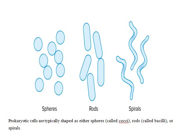
- Cells in general are small, but prokaryotic cells are really small. Typical prokaryotic cells range from in diameter, while typical eukaryotic cells range from diameterMany prokaryotic cells have sphere, rod, or spiral shapes (as shown below). In the following sections, we’ll walk through the structure of a prokaryotic cell, starting on the outside and moving towards the inside of the cell.
The capsule
Many prokaryotes have a sticky outermost layer called the capsule, which is usually made of polysaccharides (sugar polymers).
The capsule helps prokaryotes cling to each other and to various surfaces in their environment, and also helps prevent the cell from drying out. In the case of disease-causing prokaryotes that have colonized the body of a host organism, the capsule or slime layer may also protect against the host’s immune system.
Remember Griffith’s experiment, which demonstrated the existence of a “transforming principle” (DNA) that could turn rough, harmless bacteria into smooth, pathogenic bacteria? The smooth bacteria were smooth (and capable of causing disease) because they had a capsule!
The cell wall
All prokaryotic cells have a stiff cell wall, located underneath the capsule (if there is one). This structure maintains the cell’s shape, protects the cell interior, and prevents the cell from bursting when it takes up water.
The cell wall of most bacteria contains peptidoglycan, a polymer of linked sugars and polypeptides. Peptidoglycan is unusual in that it contains not only L-amino acids, the type normally used to make proteins, but also D-amino acids (“mirror images” of the L-amino acids). Archaeal cell walls don’t contain peptidoglycan, but some include a similar molecule called pseudopeptidoglycan, while others are composed of proteins or other types of polymers
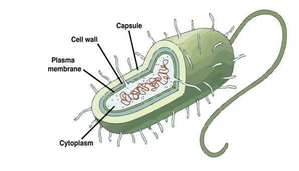
The external structures of the prokaryotic cell include a plasma membrane, cell wall, and capsule (or slime layer).
Some of the antibiotics used to treat bacterial infections in humans and other animals act by targeting the bacterial cell wall. For instance, some antibiotics contain D-amino acids similar to those used in peptidoglycan synthesis, “faking out” the enzymes that build the bacterial cell wall (but not affecting human cells, which don’t have a cell wall or utilize D-amino acids to make polypeptides)
The plasma membrane
Underneath the cell wall lies the plasma membrane. The basic building block of the plasma membrane is the phospholipid, a lipid composed of a glycerol molecule attached a hydrophilic (water-attracting) phosphate head and to two hydrophobic (water-repelling) fatty acid tails. The phospholipids of a eukaryotic or
bacterial membrane are organized into two layers, forming a structure called a phospholipid bilayer.
The plasma membranes of archaea have some unique properties, different from those of both bacteria and eukaryotes. For instance, in some species, the opposing phospholipid tails are joined into a single tail, forming a monolayer instead of a bilayer (as shown below). This modification may stabilize the membrane at high temperatures, allowing the archaea to live happily in boiling hot springs.
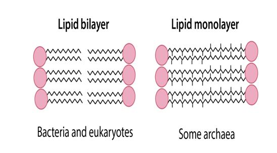
The plasma membrane of bacterial and eukaryotic (and some archael) cells is composed of a phospholipid bilayer. The tails of opposite-facing phospholipids remain separated, forming two separate layers.
The plasma membrane of some archaeal cells is composed of a phospholipid monolayer. The tails of opposite-facing phospholipids become united, forming a single layer.
Appendages
Prokaryotic cells often have appendages (protrusions from the cell surface) that allow the cell to stick to surfaces, move around, or transfer DNA to other cells.
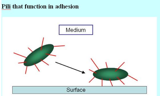
Thin filaments called fimbriae (singular: fimbria), like those shown in the picture below, are used for adhesion—that is, they help cells stick to objects and surfaces in their environment.
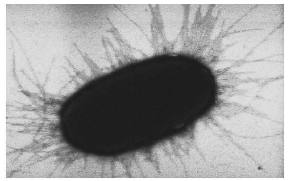
A fimbria (plural: fimbriae) is a type of appendage of prokaryotic cells. These hair-like protrusions allow prokaryotes to stick to surfaces in their environment and to each other.
Longer appendages, called pili (singular: pilus), come in several types that have different roles. For instance, a sex pilus holds two bacterial cells together and allows DNA to be transferred between them in a process called conjugation.
Another class of bacterial pili, called type IV pili, help the bacterium move around its environment.
The most common appendages used for getting around, however, are flagella (singular: flagellum). These tail-like structures whip around like propellers to move cells through watery environments.
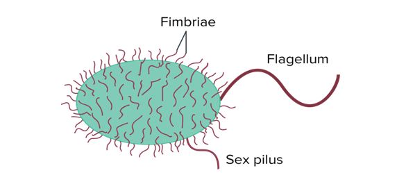
Bacteria may have various types of surface structures. These include fimbriae, short protrusions found all over the surface of the bacterium; a flagellum, found at the back of the bacterium and used for propulsion; and a sex pilus, used to grab on to other bacteria for exchange of genetic material.
Chromosome and plasmids
Most prokaryotes have a single circular chromosome, and thus a single copy of their genetic material. Eukaryotes like humans, in contrast, tend to have multiple rod-shaped chromosomes and two copies of their genetic material (on homologous chromosomes).
Also, prokaryotic genomes are generally much smaller than eukaryotic genomes. For instance, the E. coli genome is less than half the size of the genome of yeast (a simple, single-celled eukaryote), and almost times smaller than the human genome.
By definition, prokaryotes lack a membrane-bound nucleus to hold their chromosomes. Instead, the chromosome of a prokaryote is found in a part of the cytoplasm called a nucleoid.
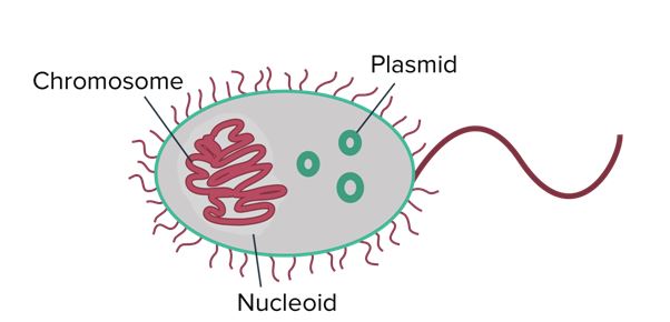
Prokaryotes generally have a single circular chromosome that occupies a region of the cytoplasm called a nucleoid. They also may contain small rings of double-stranded extra-chromosomal DNA called plasmids.
In addition to the chromosome, many prokaryotes have plasmids, which are small rings of double-stranded extra-chromosomal (“outside the chromosome”) DNA. Plasmids carry a small number of non-essential genes and are copied independently of the chromosome inside the cell. They can be transferred to other prokaryotes in a population, sometimes spreading genes that are beneficial to survival.
For instance, some plasmids carry genes that make bacteria resistant to antibiotics. (These genes are called R genes.) When the plasmids carrying R genes are exchanged in a population, they can quickly make the population resistant to antibiotic drugs. While beneficial to the bacteria, this process can make it difficult for doctors to treat harmful bacterial infections.
Cell size
Typical prokaryotic cells range from 0.1 to 5.0 micrometers (μm) in diameter and are significantly smaller than eukaryotic cells, which usually have diameters ranging from 10 to 100 μm.
The figure below shows the sizes of prokaryotic, bacterial, and eukaryotic, plant and animal, cells as well as other molecules and organisms on a logarithmic scale. Each unit of increase in a logarithmic scale represents a 10-fold increase in the quantity being measured, so these are big size differences we’re talking about!
Graph showing the relative sizes of items from, in order, atoms to proteins to viruses to bacteria to animal cells to chicken eggs to humans.
Suppose, for the sake of keeping things simple, that we have a cell that’s shaped like a cube. Some plant cells are, in fact, cube-shaped. If the length of one of the cube’s sides is , the surface area of the cube will be , and the volume of the cube will be . This means that
Eukaryotic Cell
Eukaryotic cells are defined as cells containing organized nucleus and organelles which are enveloped by membrane-bound organelles. Examples of eukaryotic cells are plants, animals, protists, fungi. Their genetic material is organized in chromosomes. Golgi apparatus, Mitochondria, Ribosomes, Nucleus are parts of Eukaryotic Cells. Let’s learn about the parts of eukaryotic cells in detail.
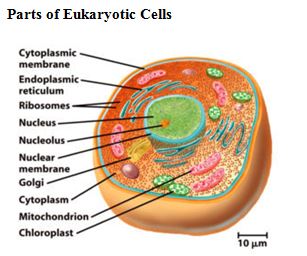
Cytoplasmic Membrane
Description: It is also called plasma membrane or cell membrane. The plasma membrane is a semi-permeable membrane that separates the inside of a cell from the outside.
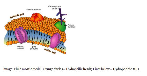
Structure and Composition: In eukaryotic cells, the plasma membrane consists of proteins, carbohydrates and two layers of phospholipids (i.e. lipid with a phosphate group). These phospholipids are arranged as follows:
-
- The polar, hydrophilic (water-loving) heads face the outside and inside of the cell. These heads interact with the aqueous environment outside and within a cell.
-
- The non-polar, hydrophobic (water-repelling) tails are sandwiched between the heads and are protected from the aqueous environments.
Scientists Singer and Nicolson described the structure of the phospholipid bilayer as the ‘Fluid Mosaic Model’. The reason is that the bi-layer looks like a mosaic and has a semi-fluid nature that allows lateral movement of proteins within the bilayer.
Functions
-
- The plasma membrane is selectively permeable i.e. it allows only selected substances to pass through.
-
- It protects the cells from shock and injuries.
-
- The fluid nature of the membrane allows the interaction of molecules within the membrane. It is also important for secretion, cell growth, and division etc.
-
- It allows transport of molecules across the membrane. This transport can be of two types:
-
-
- Active transport – This transport occurs against the concentration gradient and therefore, requires energy. It also needs carrier proteins and is a highly selective process.
-
- Passive transport – This transport occurs along the concentration gradient and therefore, does not require energy. Thus, it does not need carrier proteins and is not selective.
-
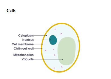
By definition, a cell is the fundamental and structural unit of all living organisms. It is the smallest biological, structural, and functional unit of all plants and animals. Therefore, cells are the ‘Building Blocks of Life’ or the ‘Basic units of Life’. Organisms made up of a single cell are ‘unicellular’ whereas organisms made up of many cells are ‘multicellular’. Cells perform many different functions within a living organism such as digestion, respiration, reproduction, etc.
For example, within the human body, a lot of cells give rise to a tissue → multiple tissues make up an organ → many organs create an organ system → several organs systems functioning together make up the human body.
Conclusion
Both DNA and RNA are made up of nucleotides, each of which contains a sugar backbone of five carbon atoms, a phosphate group, and a nitrogenous base. DNA provides the code for cellular activity, while RNA translates this code into proteins to perform cellular functions.
DNA and RNA each have four nitrogenous bases, three of which are shared (cytosine, adenine, and guanine) and one that differs between the two (RNA has uracil, while DNA has thymine). DNA stores all genetic information and helps pass it on to form new cells. The main function of RNA is to transport copies of amino acids from genes to the sites where proteins are assembled on the ribosome. This is basically done by mRNA
My Other Post-
1- Transpiration and types of Transpiration
